Download
1 / 40
460 likes | 1.06k Views
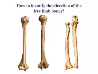
How to identify the direction of the free limb bones?. Compare the numbers and structures of upper limb bones to lower limb bones.

E N D
Compare the numbers and structures of upper limb bones to lower limb bones.
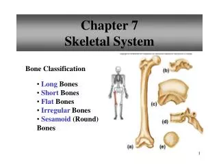
Craniumcomposed of 22 bones, divided into the calvaria (larger, upper and posterior part, contains and protects the brain),and facial skeleton(viscerocranium)(smaller, lower, and anterior part, surrounded the eye, nose and mouth). Section 5 Bones of Skull
Clavaria (8) ―Unpaired bones: frontal bone, ethmoid bone, sphenoid bone, occipital bone ―Paired bones: temporal bone parietal bone
Clavaria Frontal bone 1 Parietal bone 2 Temporal bone 2 Ethmoid bone 1 Sphenoid bone 1 Occipital bone 1
Facial skeleton (14) • Unpaired bones: mandible, vomer • Paired bones: maxilla, nasal bone, lacrimal bone, palatine bone, zygomatic bone, inferior nasal concha
parietal bone Frontal bone Sphenoid bone Temporal bone Zygomatic bone Occipital bone Nasal bone Maxilla Mandible
Squama part • Temporal bones Zygomatic prosess part External acoustic opening Mastoid part Tympanic part Mastoid process Petrous part Internal acoustic pore Styloid process
Parietal bones • Frontal bone • Occipital bone External occipital protuberance
Sphenoid bone Lesser wing Greater wing Pterygoid process Body
Ethmoid bone(on page 969) Crista galli Cribriform plate Nasal conchae Perpendicular plate Ethmoid labyrinths
Lacrimal bone Nasal bone Inferior nasal concha
Pyramidal prosess Horrizontal plate
Mandible Body of mandible • Superior border: alveolar arch • Inferior border : • Outer surface: mental foramen • Inner surface:mental spine
Rami of mandible:has two vertical processes separated by mandibular notch • Coronoid process • Condylar process • head of mandible • neck of mandible • mandibular foramen • mandibular canal • angle of mandible
Head of mandible Coronoid process Mandible Neck of mandible Mandibular foramen Mandibular notch Rami of mandible Mental foramen Angle of mandible
Neck bone • Hyoid bone Greater horn Lesser horn Body
Views of Adult Skull • Superior aspect of calvaria Bones: frontal, paired parietal, occipital Sutures: Coronal suture, Sagittal suture, Lambdoid suture • Internal aspect of calvaria sulcus for superior sagittal sinus granular foveola arterial grooves • Skull viewed from behind external occipital protuberance superior nuchal line
Internal aspect of cranial base forms three fossae • Anterior cranial fossa • Middle cranial fossa • Posterior cranial fossa
Anterior cranial fossa Formed by orbital part of frontal bone, cribriform plate of ethmoid, and lesser wings of sphenoid Structures: • crista galli • cribriform plate • cribriform foramina
Middle cranial fossa Formed by the body and greater wings of sphenoid, petrous part of temporal Structures: • hypophysial fossa • optic canal • sella turcica • carotid sulcus • superior orbital fissure • foramen rotundum
Meddle cranial fossa • foramen ovale • foramen spinosum • sulcus for middle meningeal artery • foramen lacerum • intracranial opening of carotid canal • trigeminal impression
Posterior cranial fossa Formed by occipital and the petrous part of temporal Structures: • foramen magnum • clivus • hypoglossal canal • internal occipital protuberance • groove for transverse sinus • groove for sigmoid sinus • jugular foramen • internal acoustic opening
Superior orbital fissure Cribriform plate Optic canal Hypophysial fossa Foramen rotundum Foramen spinosum Foramen ovale Foramen lacerum Internal acoustic opening Groove for sigmoid sinus Jugular foramen Hypoglossal canal Foramen magnum Groove for transverse sinus Internal occipital protuberance
Inferior view cranial base • alveolar arch • bony palate • incisive foramina • greater palatine foramen • posterior nasal apertures • foramen lacerum • jugularforamen • occipital condyle • external opening of hypoglossal canal • external opening of carotid canal • styloid process • stylomastoid foramen • mandibular fossa • articular tubercle
Incisive foramen Foramen lacerum Palatine bone Posterior nasal apertures Articular tubercle Foramen ovale Mandibular fossa Foramen spinosum Styloid process External opening of carotid canal Mastoid process Stylomastoid foramen Foramen magnum Occipital condyle
Lateral view of the skull • external acoustic opening • mastoid process • zygomatic arch • temporal fossa • pterion
Pterion External acoustic pore Zygomatic arch Mastoid process Mental foramen Condylar process Coronoid process
Anterior view Frontal region: • superciliary arch • glabella • bony orbit • bony nasal cavity
Bony orbit pyramid-shaped paired cavities • Base: supra-orbital foramen (notch), infra-orbital foramen • Apex: optic canal • Walls • Superior(roof): lacrimal fossa for lacrimal gland • Medial: lacrimal groove for lacrimal sac superior orbital fissure • Inferior(floor): inferior orbital fissure inferior orbital groove infrerior orbital canal • Lateral:
Supraorbital notch Optic canal Superior orbital fissure Lacrimal groove Inferior orbital fissure Infraorbital groove Infraorbital foramen
Bony nasal cavity • Roof: cribriform plate of ethmoid • Floor: bony palate • Lateral wall • Three nasal conchae (superior, middle and inferior) • Nasal meatus underlying each concha (superior, middle and inferior) • Spheno-ethmoidal recess above superior nasal concha • Medial wall: nasal septum • Anterior ―piriform aperture • Posterior ―posterior nasal aperture communicates with pharynx
The roof, the median septum, the floor Cribriform plate Perpendicular plate vomer Nasal cartilage maxillae Palatine bones
Superior concha Superior meatus Middle concha Middle meatus Inferior meatus Inferior nasal concha
Paranasal sinuses • Frontal sinus • Lies in frontal bone, deep to superciliary arch • Drains to anterior part of middle meatus
Maxillary sinus • Largest paired sinus, lie in the body of maxilla; • Opening into middle nasal meatus
Ethmoidal cellules • Lie in ethmoidal bone, contains large number of air cells, divided into anterior, middle and posterior groups • Anterior and middle groups drain into middle nasal meatus, while posterior group drains into superior nasal meatus • Sphenoidal sinus Lies in body of sphenoid bone • Drain into sphenoethmoidal recess
General characters of the skull at birth • The skull at birth is large in proportion to rest of the skeleton ―1/4 (adult 1/7) • The facial portion equals about one eight that of the cranium in size, whereas in adult it is one half (1/4) • Many bones consist of more than one piece • Cranial frontanelles ―unossified membrane between the bones at the angles of parietal • Anterior frontanelle ―closes during middle of 2nd year • Posterior frontanelle ―closes by the end of 6 month after birth
Highlights • Introduction • Anatomical position, terms of direction, and planes. • Osteology • Structure and classification of bones. • Formation of the bones of trunk. • The common characteristic of the vertebrae. • The main characteristic of different vertebrae. • Formation and characteristic of ribs. • Subsection of the sternum. • Definition of sternal angle and costal arch.
The primary structure of the scapula, humerus, radius and ulna. Name of the bones of hand. The primary structure of the hip bone, femur, tibia and fibula. Name of the bones of foot. The primary structure of the internal and external surface of the base of skull. The main structure of the superior, the posterior aspect and the lateral view of skull. The structure of orbit and bony nasal cavity. Names, position and opens of the paranasal sinuses.