Download
1 / 111
1.27k likes | 1.91k Views
Fluid and Electrolytes. Objectives. Identify functions of each electrolyte Identify electrolyte levels Identify signs and symptoms of each electrolyte; either hypo or hyper. Identify different buffer systems in body Identify basic blood gas values and identify abnormalities

E N D
Objectives Identify functions of each electrolyte Identify electrolyte levels Identify signs and symptoms of each electrolyte; either hypo or hyper. Identify different buffer systems in body Identify basic blood gas values and identify abnormalities Identify what happens to cells with isotonic, hypotonic, and hypertonic fluids Identify differences between whole blood, packed red blood cells, FFP, and platelets.
Fluid (Hydration) • Functions of water • Provides an extracellular transportation route to deliver nutrients to the cells and carry waste products from the cells • Provides a medium in which chemical reactions, or metabolism, can occur within the cell • Acts as a lubricant for tissues • Aids in the maintenance of acid-base balance • Assists in heat regulation via evaporation
Fluids (water) • Functions cont. • Another important influence on the amt. of water in the body is the amt. of fat on an individual • Fat contains relatively little water • The more obese an individual, the smaller the % of body water • Both obese and older adults are at risk for complications of illness from dehydration or fluid shifts d/t ↓ fluid reserve
Fluids (Water) • Percentage of body weight that is water depends on several factors. • Age • Premature infant: 90% • Newborn: 70-80% • Twelve years to adult: 50-60% • Older adults: 45-55% See Figure 22-1 p. 661 FON
Fluid Compartments • Intracellular Fluid (ICF) • Fluid contained within the cells • 2/3 to 3/4 of all body fluids are in this category • Extracellular Fluid (ECF) • Fluid contained outside the cells • Interstitial Fluid: fluid located in the spaces between the cells • Eg. Lymph, CSF, GI secretions
Fluid Compartments • Extracellular fluid cont. • Intravascular fluid: fluid that is within the blood vessels (plasma) • Plasma = water + molecules, electrolyes, proteins • (Minus blood cells and platelets)
Intake and Output • The normal daily loss of fluids must be met by the normal daily intake • Daily water intake and output is approx. 2500 ml. • Fluid leaves the body through the kidneys, lungs, skin, and GI tract • Water loss is replenished by ingestion of liquids and foods and by metabolism of food
Intake and Output • Homeostasis • “Relative constancy in the internal environment” • Naturally maintained by adaptive responses healthy survival • BALANCE • Intake includes: all fluids entering the body • Output includes: all fluids leaving the body
Intake and Output • Output includes: • Urine • Diarrhea • Vomitus • Ng suction • Drainage bags/wound drainage • Insensible loss: perspiration, expiration
Intake and Output • Role of the Kidneys in fluid balance • Proper functioning of the kidneys healthy regulation of fluid balance • Nephrons (kidney cells) filter blood urine output • Kidneys must excrete a minimum of 30ml/hr of urine in order to eliminate waste products from the body
Intake and Output • If the body loses 1-2% of its fluid: • Kidneys conserve fluid by reabsorbing more water from the renal filtrate concentrated urine • If there is excess body fluid, the kidneys excrete more urine and it is more dilute • This rids the body of excess fluid and conserves electrolytes
Intake and Output • Nursing Implications: • I and O Record • Daily weight • Same time • Same amt. of clothing and/or bed linens • Same equipment • Empty all drainage bags • Checking urine Specific Gravity • Nml range: 1.010 1.030 • < 1.010 = dilute urine • > 1.030 = concentrated urine
Intake and Output • Note: 1 L of fluid = 2.2 lbs (1 kg.) • A weight change of 2.2 lbs will reflect a loss or gain of 1 L body fluid
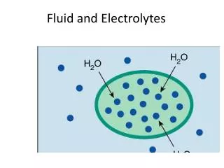
Movement of Fluid and Electrolytes To carry out cellularfunctions, substancesmust cross the semipermeable membrane surrounding each body cell to enter the cell. The fat and protein molecules that make up the membrane are arranged so that some substances can enter the cells and others cannot.
Solutions • Isotonic, hypotonic, hypertonic • “Iso” – “the match” • Has the same solution concentration as another solution • Ex. : normal saline solution – the concentration of sodium in the solution nearly equals the concentration of sodium in the blood.
Isotonic fluids Can be helpful in hypotensive or hypovolemic patients. Can be harmful. There is a risk of fluid overloading, especially in patients with CHF and hypertension.
Solutions • “hypotonic” solution: • Has a lower solute concentration than another solution • Ex. “0.5 % NS” is considered hypotonic because the concentration of sodium in the solution is lower than the concentration of sodium in the pts. blood • Water is pulled from the vascular compartment into the interstitial fluid compartment. Then, as the interstitial fluid is diluted, this assists in the transpor- tation of water into the adjacent cells.
Hypotonic fluids Can be helpful when cells are dehydrated such as a dialysis patient on diuretic therapy. May also be used for hyperglycemic conditions like diabetic ketoacidosis, in which high serum glucose levels draw fluid out of the cells and into the vascular and interstitial compartments.
Solution • “Hypertonic”: • Has a higher solute concentration than another solution- for our uses- blood • Ex. A solution of D5W 0.9%NS is considered hypertonic because the concentration of solution in the solution is greater than the concentration of solutes in the patient’s blood.
Hypertonic Solution • Pulls fluid and electrolytes from the intracellular and interstitial compartments into the intravascular compartment. • Can help stabilize blood pressure, increase urine output, and reduce edema. • Used in head trauma to decrease swelling of the brain. • Care must be taken with their use. Dangerous in the setting of cell dehydration.
Movement • A number of processes allow this mass movement of substances into and out of cells • Transport Processes = • Passive Transport • Active Transport
Movement • Active Transport Process • Chemical substance called “ATP” (adenosine triphosphate) is produced in cells • Releases energy cell able to work • For active transport to occur, need: • Breakdown of ATP + use of related (cell) energy • Substances actively transported through the cellular membrane includes: • Na⁺, K⁺, Ca⁺⁺, Fe⁺, H⁺ and amino acids • Glucose
Movement • Passive Transport • Movement of small molecules by diffusion across a cell membrane • No cellular energy required to move substances from a ↑ concentration to a ↓concentration • Active Transport • Cellular energy is required to move substances from ↓ concentration ↑ concentration
This is the movement of water from an area of lower solute concentration to an area of higher concentration. Solutes move from an area of higher concentration to an area of lower concentration, which eventually results in an equal distribution of solutes within the two areas. This is the transfer of water or dissolvedsubstances from an area of higher pressure to an area of lower pressure. • Passive Transport Processes • No cellular energy is required to move substances from a high concentration to a low concentration. • Osmosis: • Diffusion: • Filtration:
Movement • Filtration: • Ex.: at the capillary level of the circulation • Force behind filtration: hydrostatic pressure = the force of fluid pressing outward on a vessel wall • The pumping action of the heart is responsible for the amount of force that causes water and electrolytes to move from the capillaries to the interstitial fluid.
Electrolytes • As water moves through the compartments of the body, it contains substances that are sometimes called “minerals” or “salts” • These are technically known as “electrolytes” • Electrolytes develop tiny electrical charges when they dissolve in water and break up into particles known as ions.
Electrolytes • Ions develop either a positive or negative electrical charge. • Cations have a positive charge. • Anions have negative charge. • A balance exists between the electrolytes; for each positively charged cation, there must be a negatively charged anion. • Chemical designations: Na+, K+, Cl-, Ca++
Electrolytes • Measured by their electrical activity • To measure: blood sample drawn • Expressed as “milliequivalents” – mEq • A mEq refers to the combining power of an ion
Sodium (Na⁺) • Most abundant electrolyte in the body • Normal level: 134 to 142 mEq/L • Major source is the diet; frequently must be limited • Functions of sodium: • regulates water balance, • controls extracellular fluid volume – water follows sodium in the body • increases cell membrane permeability, • stimulates conduction of nerve impulses, • helps maintain neuromuscular irritability, • controls contractility of muscles
Hyponatremia • Less than normal concentration of sodium in the blood • Caused by a sodium loss or a water excess (e.g. excessive hydration) • With the lower sodium, the body’s compensation results in decreased water excretion • Dilution of the extracellular fluid, causes a fluid shift into the cells, which in turn causes swelling. The brain is the most susceptible.
Signs and Symptoms of Hyponatremia Headache, fatigue, Postural Hypotension Muscle weakness, twitching or tremors Apathy, lethargy Nausea & Vomiting Abodominal cramping Death by shock or coma
Remember , Fluid excess or Sodium loss or Both She had successfully completed a marathon 2 years prior to running in Boston. By all accounts she looked great all throughout the 19 miles of the 2002 Boston Marathon. Yet, she became unsteady and collapsed between miles 19 and 20. She was placed in an ambulance and had a seizure on her way to the hospital. She never regained consciousness. Since Cynthia's passing there have been several reports intended to warn athletes about the dangers of hyponatremia so that runners can be better informed.
Hypernatremia Greater than normal concentration of sodium in the blood Can occur when there is a sodium excess or a water loss (dehydration) Body attempts to correct the imbalance by conserving water through renal reabsorption Causes fluid to shift from the cells (high concentration) to the interstitial spaces (low concentration), resulting in cellular dehydration
Signs and Symptoms ofHypernatremia • Dry, tenacious mucous membrane • Reduced and concentrated urine output • A decrease in the turgor of the skin which might appear as a sunken dark look to the skin around the eyes • Restlessness, agitation, confusion, flushed skin • Tachycardia, capllary refill time (>2 seconds), even cyanosis, mottling, and reticulation
Signs and Symptoms of Hypernatremia • Weight loss • Principal manifestations involve the CNS - confusion, altered mental status, obtundation, stupor, coma, increased neuromuscular irritability (twitching, seizures) • Intracerebral and subarachnoid hemorrhage may result if shrinkage of brain volume leads to tearing of the bridging veins. • Death by cardiac arrythmias
Potassium (K⁺) • Normal level is 3.5 to 5 mEq/L • Well-balanced diet usually provides adequate potassium. • The kidneys control the excretion of potassium. • Na⁺ and K⁺ seem to pair off against each other and the kidneys prefer to conserve Na⁺ and excrete K⁺ • Any condition that causes a decrease in urine output K⁺ retention
Potassium (K⁺) • Function of potassium • Main function is regulation of water and electrolyte content within the cell. • Required for nerve conduction, muscle function • Involved in cellular enzyme activities • Role in acid-base balance: control hydrogen ion concentration
Hypokalemia Decrease in body’s potassium to a level below 3.5 mEq/L The major cause of loss is renal excretion (diuretics). The kidneys do not conserve potassium and excrete it even when the body needs it. Potassium can be depleted due to excessive GI losses from gastric suctioning or vomiting and the use of diuretics. Affects skeletal and cardiac function.
Signs and Symptoms of Hypokalemia Skeletal muscle weakness especially in lower legs; leg cramps Cardiac dysrhythmias. Orthostatic hypotension Decreased bowel sounds, cramps, constipation, anorexia, Nausea and vomiting. Hyporeflexia Numbness, tingling, Paresthesia Extreme weakness of the body with “flaccid paralysis” or limpness. Paralysis of the muscles of the lungs results in death. Abnormal heart beat (arrhythmia) that can lead to death from cardiac arrest
Hyperkalemia Increase in the body’s serum potassium level above 5 mEq/L The major cause of excess potassium is renal disease or ingestion, orally or IV Severe tissue damage causes potassium to be released from the cell. Excessive increase in foods high in potassium can cause serum levels to increase.
Signs and Symptoms of Hyperkalemia • EKG changes • Irregular heartbeat; hypotension • Difficulty breathing • Nausea & Vomiting • Diarrhea, colic • Skeletal muscle weakness • Tingling, numbness, or other unusual sensations • Flaccid paralysis
Hyperkalemia • The primary cause of morbidity and death is potassium's effect on cardiac function. • The mortality rate can be as high as 67% if severe hyperkalemia is not treated rapidly.
Chloride (Cl⁻) • Normal level is 96 to 105 mEq/L. • It is the chief anion in interstitial and intravascular fluid. • It has the ability to diffuse quickly between the intracellular and extracellular compartments and combines easily with sodium to form sodium chloride (Na⁺Cl⁻) or with potassium to form potassium chloride (K⁺Cl⁻) • The main route of excretion is the kidneys
Chloride (Cl⁻ Hypochloremia Hyperchloremia Poss. when bicarbonate level falls ↑ in Cl⁻ ions Chloride imbalances rarely occur independently of other electrolytes no s/sx to identify a Cl⁻ imbalance • Usually occurs when Na⁺ is lost • (Na⁺ and Cl⁻) are frequently paired) • From: • Vomiting • Prolonged Ng suctioning • Fistula drainage
Calcium (Ca⁺⁺) • Normal level is 4.5 mEq/L. • Dietary Intake is source especially milk and cheese • Calcium is deposited in the bones, and • Mobilized as needed to help keep the blood level constant during any period of insufficient intake. • Vitamin D, calcitonin, and parathyroid hormone are necessary for absorption and utilization of calcium.
Calcium (Ca⁺⁺) • 3 considerations are important in blood calcium level: • 1. deposition and resorption of bone • 2. absorption of calcium from GI tract • 3. excretion of calcium in urine and feces