Chapter 8 The Appendicular Skeleton
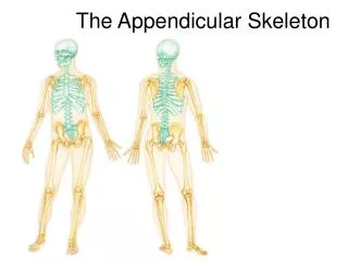
Chapter 8 The Appendicular Skeleton. Course objectives: List the bones of the appendicular skeleton Describe and identify the bones of the pectoral girdle Describe and identify the bones of the pelvic girdle. Appendicular Skeleton.

Chapter 8 The Appendicular Skeleton
E N D
Presentation Transcript
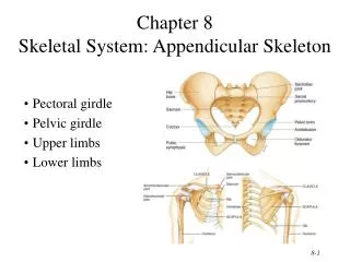
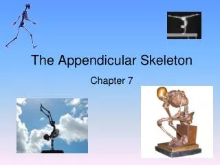
Chapter 8 The Appendicular Skeleton Course objectives: List the bones of the appendicular skeleton Describe and identify the bones of the pectoral girdle Describe and identify the bones of the pelvic girdle
Appendicular Skeleton • Includes the bones of the upper limb and their attachments to the axial skeleton at the pectoral girdle. • Includes the bones of the lower limb and their attachments to the axial skeleton at the pelvic girdle.
Pectoral Girdle • scapula – “shoulder blade” -(triangular flat bone) articulates with humerus of arm at the glenoid fossa • clavicle – “ collar bone" -flat bone articulates with the acromion process of scapula and the manubrium of the sternum, thus forming the only bony link with the axial skeleton and pectoral appendicular skeleton
Scapula • Thin triangular flat bone that forms the bulk of the shoulder • Connects to the arm at the humerus via glenoid fossa • Connects to the clavicle at the acromion process
Scapula landmarks • Supraspinous and infraspinous fossa • Suprascapular fossa • Acromion • Coracoid process • Glenoid cavity • Lateral and medial border
Clavicle landmarks • Acromial end • Sternal end • Conoid tubercle • Costoclavicular tuberosity
The Upper Limb • Consists of 30 bones • Grouped into bones of the arm, forearm and hand • Arm = Humerus • Forearm = Radius and Ulna • Hand = Carpals (8), metacarpals (5) and phalanges (5)
Humerus landmarks • Head and body of humerus • Greater and Lesser tubercles • Anatomical neck and Surgical neck • Medial and lateral supracondylar ridges • Medial and lateral epicondyle • Olecranon and radial fossa • Coronoid process • Deltoid tuberosity • Capitulum • Trochlea
Forearm “antebrachium” • Consists of the Radius (lateral) and Ulna (medial). • Both are connected along their length by a ligament (interosseous membrane)
Radius landmarks • Head, neck and shaft • Radial tuberosity • Ulnar notch • Styloid process • Nutrient foramen
Ulna landmarks • Olecranon process • Coronoid process • Trochlear notch • Radial notch • Head of the ulna • Styloid process of ulna
The Hand • Consists of: • Carpals (8) “wrist” • Metacarpals (5) “palm” • Phalanges (5) “fingers”
Carpal bones Eight bones makeup the wrist
Metacarpals and Phalanges • These bones are not named individually but are numbered 1-5. • The thumb “pollex” is number 1. • They are all long bones • The base of the metacarpals articulate with the carpal bones at their base and the phalanges at their head. • The phalanges consist of a proximal, middle and distal phalanx in all but the thumb .
The Pelvic Girdle • The “hips” forma much more solid and stable connection for the lower limbs to the axial skeleton than the pectoral girdle is to the upper limbs. • The pelvic girdle is formed by the coxal bones (a.k.a. hip bones, os coxae) which fuse posteriorly with the sacrum. • The coxal bones are formed by the fusion of three separate bones ( ilium, ischium and pubis) during growth.
“Os Coxae” Hip bones • Formed by the fusion of three bones -1. ilium, 2. ischium, and 3. pubis • Attaches to the lower limb and spine at sacroiliac joint • Supports the pelvic organs or viscera • Attached to the axial skeleton by strong ligaments
Os coxae landmarks • Iliac crest • Anterior superior and ant. inferior iliac spine • Posterior superior and post. inferior iliac spine • Greater and lesser sciatic notch • Iliac fossa • Ischial spine and tuberosity; ramus of ischium • Obturator foramen • Superior and inferior ramus of pubis • Pubic symphysis and pubic arch • Acetabulum
Male vs Female Os coxae • Significant differences exist between the male and female pelvis. • ♀ pelvic outlet is enlarged due to in part greater separation of ischial spines • ♀ less curvature of sacrum and coccyx which in males ♂ arcs into pelvic outlet • ♀ wider more circular pelvic inlet • ♀ relatively broad, low pelvis • A broader pubic angle in ♀ between pubic bones > 100°
True vs. False pelvis • False pelvis = area within entire pelvic girdle • True pelvis = area below pelvic brim
Lower limb Consists of: • Femur “Thigh” = hip to the knee • Tibia and fibula “Leg” = knee to foot • Foot
Femur “Thigh” landmarks • Longest, strongest, largest bone in body • Head • Neck • Greater and lesser trochanter • Medial and lateral condyle • Medial and lateral epicondyle • Linea aspera
Patella “knee cap” landmarks • Base • Apex • Articular surfaces
Lower leg • technically the distance between the knee and ankle • Bones of the leg: tibia (shin bone) and fibula (lateral leg bone) • Consists of the: Tibia (shin bone) Fibula • Interosseuos membrane connects tibia and fibula along their length
Tibia landmarks • Medial and lateral condyle • Tibial tuberosity • Medial malleolus (medial bulge of ankle) • Anterior border (crest) is the shin
Fibula landmarks • Fibula is lateral bone of the leg • Head • Lateral malleolus (lateral bulge of ankle)
The foot • Includes the bones of the; -Tarsus -Metatarsus Phalanges • Functions -support of the body -lever for walking or running
Tarsal bones • Talus • Calcaneous
Metatarsals and Phalanges • Are all long bones • Metatarsals numbered 1-5 • Phalanges consist of proximal, middle and distal bones in all but big toe • Big toe or great toe is Hallux