The Appendicular Skeleton
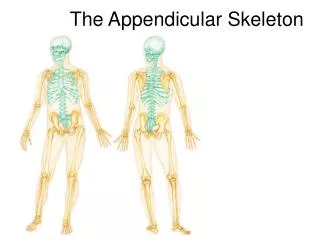
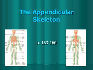
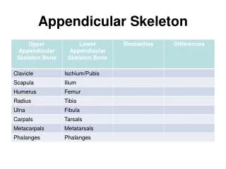
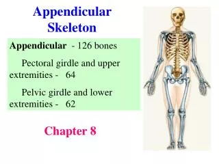
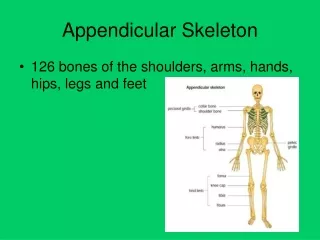
The Appendicular Skeleton. The Appendicular Skeleton. 126 bones Limbs (appendages) Pectoral girdle (attaches arm to the axial skeleton) Pelvic girdle (attaches leg to the axial skeleton). The Pectoral (Shoulder) Girdle. Composed of two bones Clavicle - collarbone

The Appendicular Skeleton
E N D
Presentation Transcript
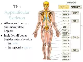
The Appendicular Skeleton • 126 bones • Limbs (appendages) • Pectoral girdle (attaches arm to the axial skeleton) • Pelvic girdle (attaches leg to the axial skeleton)
The Pectoral (Shoulder) Girdle • Composed of two bones • Clavicle - collarbone • Scapula – shoulder blade
The Pectoral (Shoulder) Girdle • These bones are very light and allow the upper limb to have exceptionally free movement because: • Each pectoral girdle attaches to axial skeleton at only 1 point = sternoclavicular joint • The loose attachment of the scapula allows it to slide back & forth against the thorax • The glenoid cavity is shallow & the shoulder joint is poorly reinforced by ligaments
Bones of the Shoulder Girdle Figure 5.20a–b
The Pectoral (Shoulder) Girdle • Consists of 2 bones • Clavicle (collar bone) = slender, double curved bone • Attaches to manubrium of the sternum medially & to the scapula laterally • Acts as a brace to hold the arm away from the thorax & helps prevent shoulder dislocation
The Pectoral (Shoulder) Girdle • Scapula (shoulder blade) = triangular & flare when we move our arms posteriorly (“wings”) • Not directly attached to axial skeleton; held in place by trunk muscles • Flattened body w/ 2 processes: • Acromion process • Coracoid process
The Pectoral (Shoulder) Girdle • Acromion process = the enlarged end of the spine of the scapula • Connects w/ the clavicle @ the acromioclavicular joint • Coracoid process = beaklike • Points over the shoulder & anchors some of the arm muscles • Suprascapular notch serves as a nerve passageway
The Pectoral (Shoulder) Girdle • Scapula has 3 borders & 3 angles: • Superior • Medial (vertebral) borders • Lateral (axillary) • Superior • Inferior Angles • Lateral
Bones of the Shoulder Girdle Figure 5.20c–d
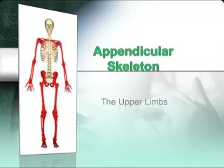
Bones of the Upper Limb (30) • The arm is formed by a single bone • Humerus • Rounded proximal end fits into glenoid cavity of scapula Figure 5.21a–b
Bones of the Upper Limb (30) • Opposite of the head – 2 bony processes = greater & lesser tubercles, sites of muscle attachment • Midpoint of shaft – deltoid tuberosity where the deltoid m. attaches Figure 5.21a–b
Bones of the Upper Limb (30) • Radial groove runs obliquely down the posterior shaft • Distal end – medial trochlea (looks like a spool) & lateral ball-like capitulum – both articulate w/ bones of forearm Figure 5.21a–b
Bones of the Upper Limb (30) • Above the trochlea anteriorly is a depression – coronoid fossa • On posterior surface is the olecranon fossa • Both are flanked by medial & lateral epicondyles Figure 5.21a–b
Bones of the Upper Limb • The forearm consists of the radius and the ulna. • Radius is lateral when the arm is in the anatomical position (on the thumb side) & the ulna is medial • When the hand is rotated, the distal end of the radius crosses over & ends up medial to the ulna. Figure 5.21c
Bones of the Upper Limb • Radius & ulna articulate @ small radioulnar joints • They are connected along their entire length by a flexible interosseous membrane • Both have a styloid process @ their distal end Figure 5.21c
Bones of the Upper Limb • Disc-shaped radial head forms a joint w/ the capitulum of the humerus. • Below the head is the radial tuberosity where tendon of biceps m. attaches. Figure 5.21c
Bones of the Upper Limb • On the ulna’s proximal end are the anterior coronoid process & the posterior olecranon process, which are separated by the trochlear notch. • These 2 processes grip the trochlea of the humerus in a pliers-like joint. Figure 5.21c
Bones of the Upper Limb • Hand consists of carpals, metacarpals & phalanges • 8 carpals arranged in 2 irregular rows of 4 bones each form the part of the hand called the carpus (wrist) • Bound together by ligaments that restrict movements between them. Figure 5.22
Bones of the Upper Limb • Metacarpals form the palm • Numbered 1 to 5 from the thumb to the pinky • Clenched fist – heads of the metacarpals = knuckles • Phalanges (14) are the finger bones (3 in each finger & 2 in the thumb) Figure 5.22
Bones of the Upper Limb • Carpals starting Left to Right (pinky to thumb - both rows): • Joe took a Hamate and hit poor Pete in the Capitate, breaking it into a Trapezoid and Trapezium. He did it b/c he was Pisiform(d) Triquetral times. The Judge said he was Lunate and sent him to the Scaphoid. Figure 5.22
The Bony Pelvis vs. the Pelvic Girdle • Bony Pelvis • Composed of: • Sacrum • Coccyx • Coxal bones (coxae) or ossa coxae (Hip bones) • Pelvic Girdle • Coxae are composed of 3 pair of fused bones • Ilium • Ischium • Pubic bone
The Bony Pelvis Figure 5.23a
The Pelvic Girdle: Right Coxal Bone Figure 5.23b
The Pelvic Girdle • Coxae are large, heavy & attached securely to the axial skeleton. • Sockets that receive the femur are deep & heavily reinforced w/ ligaments. • Function = bearing weight; total wt. of upper body rests on pelvis. • Reproductive organs, bladder & part of large intestine lie within & are protected by pelvis.
The Pelvic Girdle • Each coxais formed by the fusion of 3 bones: • Ilium (Large flaring bone - forms most of the coxa) • Connects posteriorly w/ sacrum @ the sacroiliac joint. • Alae - winglike portions of the ilia. • Iliac crest – upper edge of alae that ends anteriorly in the anterior superior iliac spine & posteriorly in the posterior superior iliac spine w/ small inferior spines located below these.
The Pelvic Girdle • Ischium (“sit down bone”) • Most inferior part of coxa. • Ischial tuberosity, a roughened area, receives body wt. when sitting. • Ischial spine, superior to the tuberosity, narrows the outlet through which the baby passes during childbirth. • Greater sciatic notch allows blood vessels & the large sciatic nerve to pass from the pelvis posteriorly into the thigh.
The Pelvic Girdle • Pubis or pubic bone • Most anterior part of the coxa. • Fusion of the rami of the pubis anteriorly & the ischium posteriorly forms a bar of bone enclosing the obturator foramen, an opening through which blood vessels & nerves pass into the anterior part of the thigh. • Pubic bones fuse anteriorly to form a cartilaginous joint called the pubic symphysis.
The Pelvic Girdle • The ilium, ischium, & pubis fuse @ the deep socket called the acetabulum (“vinegar cup”); it receives the head of the femur.
The Bony Pelvis • Bony pelvis is divided into 2 regions: • False pelvis, superior to the true pelvis, is the area medial to the flaring portions of the ilia. • True pelvis lies inferior to the flaring parts of the ilia & the pelvic brim.
The Bony Pelvis • Dimensions of the true pelvis are important for childbirth – must be large enough for the head to pass. • Outlet is the inferior opening of the pelvis. • Inlet is the superior opening.
Differing Characteristics Between the Male and Female Pelvis • Female inlet is larger & more circular. • Female pelvis as a whole is shallower & the bones are lighter & thinner. • Female ilia flare more laterally.
Differing Characteristics Between the Male and Female Pelvis • Female sacrum is shorter & less curved. • Female ischial spines are shorter & farther apart; thus the outlet is larger. • Female pubic arch is more rounded because the angle of the pubic arch is greater.
Bones of the Lower Limbs • Carry our total body weight when standing = thicker & stronger • The thigh has one bone – Femur (thigh bone) Figure 5.24a–b
Bones of the Lower Limbs • Femur (thigh bone) • Heaviest, strongest bone in the body. • Proximal end has ball-like head, neck and greater & lesser trochanters. Trochanters are separated anteriorly by intertrochanteric line and posteriorly by intertrochanteric crest. Figure 5.24a–b
Bones of the Lower Limbs • Trochanters, inter.-crest & gluteal tuberosity are sites for muscle attachment. • Slants medially as it runs downward to bring knees in line w/ body’s center of gravity. (more noticeable in females b/c of wider pelvis) Figure 5.24a–b
Bones of the Lower Limbs • Distally are the lateral & medial condyles – articulate w/ tibia. (condyles separated by condylar fossa) • Anteriorly on distal end is the patellar surface – forms a joint w/ patella (kneecap) Figure 5.24a–b
Bones of the Lower Limbs • The leg has two bones – tibia and fibula • Connected by interosseous membrane Figure 5.24c
Bones of the Lower Limbs • Tibia (shinbone) • Larger & more medial • At proximal end – medial & lateral condyles (separated by intercondylar eminence) articlulate w/ distal end of femur to form knee joint. Figure 5.24c
Bones of the Lower Limbs • Patellar ligament attaches to tibial tuberosity (anter.) • Distally, a process called medial malleolus forms inner bulge of ankle. • Anterior surface has sharp ridge – anterior border (unprotected by muscle – so you can feel this) Figure 5.24c
Bones of the Lower Limbs • Fibula • Thin & sticklike • Distally - lateral malleolus forms outer part of ankle. Figure 5.24c
Bones of the Lower Limbs • Foot – composed of tarsals, metatarsals & phalanges • Two important fxns.: supports body weight & serves as a lever allowing us to propel body forward when walking,etc. Figure 5.25
Bones of the Lower Limbs • 7Tarsalbones • Weight carried by the 2 largest tarsals: • calcaneus (heelbone) & • talus (lies b/n tibia & calcaneus) Figure 5.25
Bones of the Lower Limbs • 5 metatarsals form the sole • 14 phalanges form the toes (each toe has 3, except the big toe) Figure 5.25
Arches of the Foot • Bones are arranged to form 3 strong arches: • 2 longitudinal (medial & lateral), 1 transverse • Ligaments bind foot bones together • Tendons of the foot muscles help to hold bones in arched position but allow for “springiness” – weak arches are referred to as “fallen arches” or “flat feet” Figure 5.26