Chapter 5 Viruses
Chapter 5 Viruses. Chapter outline. 5.1 General Properties of Viruses 5.2 General Features of Virus Reproduction 5.3 Overview of Bacterial Viruses 5.4 Temperate Bacteriophages: Lysogeny and Lambda 5.5 Overview of Animal Viruses 5.6 Pox Viruses 5.7 Adcnoviruses

Chapter 5 Viruses
E N D
Presentation Transcript
Chapter outline 5.1 General Properties of Viruses 5.2 General Features of Virus Reproduction 5.3 Overview of Bacterial Viruses 5.4 Temperate Bacteriophages: Lysogeny and Lambda 5.5 Overview of Animal Viruses 5.6 Pox Viruses 5.7 Adcnoviruses 5.8 Retroviruses 5.9 Viroids and Prions
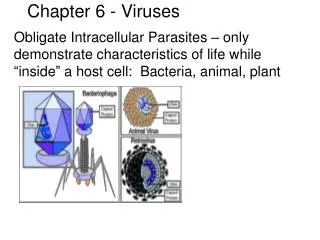
Concepts • Viruses are simple, acellular entities consisting of one or more molecules of either DNA or RNA enclosed in a coat of protein. They are reproduced only within living cells and are obligately intracellular parasites • The nucleic acid strands can be linear, closed cycle, or able to assume either shape. • Viruses are classified on the bases of their nucleic acid’s characteristics, capsid symmetry, the presence or absence of an envelop, their host and other properties.
5.1 General Properties of Viruses Viruses differ from living cells in at least three ways: (1) Their simple, acellular organization , (2) The absence of both DNA and RNA in the same virion, (3) Their inability to reproduce independently of cells and carry out cell division as prokaryotes and eukaryotes do.
Viruses can exist in two phases Extracellular and intracellular Virion, the extracellular phase, posses few if any enzymes and can not reproduce independently of living cells. In the intracellular phase, viruses exist primarily as replicating nucleic acids that induce host metabolism to synthesize virion components; eventually complete virus particles or virions are released.
Hosts The particular host range of a virus is determined by the virus's requirements for its specific attachment to the host cell and the availability within the potential host of cellular factors required for viral multiplication. Three main classes - animal viruses, bacterial viruses (bacteriophages), and plant viruses.
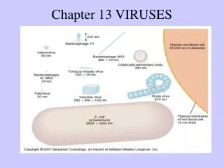
Size Viruses vary considerably in size. Although most are quite a bit smaller than bacteria, some of the larger viruses (such as the smallpox virus) are about the same size as some very small bacteria (such as the mycoplasmas, rickettsias, and chlamydias). Viruses range from 20 to 300 nm in diameter
Virus particles (virions) vary widely in size and shape. Viruses are smaller than cells, ranging in size from 0.02 to 0.3 um. Smallpox virus, one of the largest viruses, is about 200 nm in diameter; poliovirus, one of the smallest, is only 28 nm in diameter.
The genomes of viruses can be composed of either DNA or RNA, and some use both as their genomic material at different stages in their life cycle. However , only one type of nucleic acid is found in the virion of any particular type of virus.
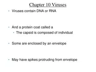
Structure of viruses • Most viruses are too small to be seen under light microscope. • All viruses consists of an RNA or DNA core genome surrounded by a protein coat capsid. • The combined viral genome and capsid is called the nucleocapsid.
Complex viruses Some viruses have complicated structures and are called complex viruses. Examples of complex viruses are poxviruses, which do not contain clearly identifiable capsids but have several coats around the nucleic acid. Certain bacteriophages have capsids to which additional structures are attached.
General morphology Viruses may be classified into several morphological types on the basis of their capsid architecture as revealed by electron microscopy and a technique called x-ray crystallography.
A.Some viruses, such as tobacco mosaic virus, have a helical symmetry with the capsid surrounding an RNA genome. B.Many viruses that infect bacteria, such as the T-even bacteriophage, have a complex capsid with DNA contained within the head structure.
C.Some animal viruses, such as adenovirus, have isometric symmetry and a DNA genome. D.Others, such as coronavirus, have complex capsids and an envelope with protruding proteins surrounding an RNA genome.
Helical viruses Helical viruses resemble long rods that may be rigid or flexible. Surrounding the nucleic acid, their capsid is a hollow cylinder with a helical structure. An example of a helical virus that is a rigid rod is the tobacco mosaic virus.
Polyhedral viruses The capsid of most polyhedral viruses is in the shape of a regular polyhedron with 20 triangular faces and 12 corners. The capsomeres of each face form an equilateral triangle. An example of a polyhedral virus is the adenovirus. Another is the poliovirus.
Enveloped viruses The capsid of viruses is covered by an envelope. Enveloped viruses are roughly spherical but variable in shape. When helical or polyhedral viruses are enclosed by envelopes, they are called enveloped helical and enveloped polyhedral viruses.
5.2 General Features of Virus Reproduction The virus must induce a living host cell to synthesize all the components needed to make virus particles. These components must then be assembled into the proper structure, and the new virions must escape from the cell and infect other cells. The phases of this replication process in a bacteriophage can be categorized in seven steps.
1. Attachment 2. Penetration 3. Early steps in replication 4. Replication 5. Synthesisof proteins 6. Assembly 7. Release
Multiplication of bacteriophages 1. Attachment (adsorption) of the virion to a susceptible host cell. 2. Penetration (injection) of the virion or its nucleic acid into the cell. 3. Early steps in replication during which the host cell biosynthetic machinery is altered as a prelude to virus nucleic acid synthesis. Virus-specific enzymes are typically made.
4. Replication of the virus nucleic acid. 5. Synthesis of proteins used as structural subunits of the virus coat. 6. Assembly of structural subunits (and membrane components in enveloped viruses) and packaging of nucleic acid into new virus particles. 7. Release of mature virions from the cell.
Attachment of phage to host cell : After a chance collision between phage particles and bacteria, an attachment site on the virus attaches to a complementary receptor site on the bacterial cell.
Penetration: During the process of penetration, the bacteriophage's tail releases an enzyme, phage lysozyme, which breaks down a portion of the bacterial cell wall. then the bacteriophage injects its DNA (nucleic acid) into the bacterium.
Biosynthesis of viral components: Any RNA transcribed in the cell is mRNA transcribed from phage DNA for biosynthesis of phage enzymes and capsid protein. The host cell's ribosomes, enzymes, and amino acids are used for translation.
Formation of mRNA after infection of cells by viruses of different types. The chemical sense of the mRNA is considered as plus(+). The sense of the various virus nucleic acids are indicated as+if the same as mRNA, as–if opposite, or as+–if double–stranded. Examples are indicated next to the virus nucleic acid.
The phage heads and tails are separately assembled from protein subunits, the head is packaged with phage DNA, and the tail is attached. Maturation and release:
Release: Lysozyme, whose code is provided by a phage gene, is synthesized within the cell. This enzyme causes a breakdown of the bacterial cell wall, and the newly produced bactedophages are released from the host cell. The number of newly synthesized phage particles released from a single cell usually ranges from about 50 to 200.
Following adsorption, the infectivity of the virus particles disappears, a phenomenon called eclipse. This is due to the uncoating of the virus particles. During the latent period, replication of viral nucleic acid and protein occurs. The maturation period follows, when virus nucleic acid and protein are assembled into mature virus particles. At this time, if the cells are broken up, active virus can be detected. Finally, release occurs, either with or without cell lysis.
The liming of the one-step growth cycle varies with the virus and host. With many bacterial viruses, the whole cycle may be complete in 20-60 min, whereas with animal viruses 8-40 hr is usually required for a complete cycle.
Eclipse period • There are genetic controls that regulate when different regions of phage DNA are transcribed into mRNA during the multiplication cycle. • There are early messages that are translated into early phage proteins, the enzymes used in the synthesis of phage DNA.
There are late messages that are translated into late phage proteins for the synthesis of capsid proteins. • This control mechanism is mediated by RNA polymerase.
Following injection of DNA, early and middle mRNA is produced that codes for nucleases, DNA polymerase, new phage-specific sigma factors, and various other proteins involved in DNA replication. Late mRNA codes for structural proteins of the phage virion and for T4 lysozyme, needed to lyse the cell and release new phage particles.
5.3 Overview of Bacterial Viruses Most of the bacterial viruses that have born studied in detail infect bacteria of the enteric group, such as Escherichia coli and Salmonella typhimurium. However, viruses are known that infect a variety of prokaryotes, both bacteria and archaea. A few bacterial viruses have lipid envelopes but most do not. However, many bacterial viruses are structurally complex, with head and complex tail structures.
Schematic representations of the main types of bacterial viruses The structures of M13, Φχ174, MS2, T4, lambda, T7 and Mu. sizes are to approximate scale. Many viruses are structurally complex, with head and complex tail structures. There is also a great diversity in the manner in which virus multiplication occurs.
Head Tail Tail fiber Endplate Note the complex structure. The tail components are involved in attachment of virion to the host and infection of the nucleic acid the head is about 85 nm in diameter
The bacterial RNA viruses are all quite small, about 26 nm in size, and they are all icosahedral, with 180 copies of coat protein per virus particle. The complete nucleotide sequences of several RNA phage genomes are known. The genome of the RNA phage MS2, which infects Escherichia coli, is 3569 nucleotides long. The RNA strand in the virion acts directly as mRNA on entry into the cell.
Flow of events during viral multiplication The small genome encodes only four proteins. These are the maturation protein, coat protein, lysis protein, and a subunit of RNA replicase, the enzyme that brings about replication of the viral RNA. The RNA replicase is a composite protein, composed of the virus-encoded polypeptide and host polypeptides. The virus appears to employ host proteins that have distinct functions and use them to make viral replicase.
Quantification of bacterial virus by plaque assay 2. The mixture poured on the surface of a nutrient agar plate. 3. The host bacteria begin to grow, and after overnight incubation form a lawn of confluent growth. 1. A dilution of a suspension containing the virus material is mixed in a small amount of melted agar with the sensitive host bacteria.
Phage plaques Photograph of a plate showing plaques formed by bacteriophage on a lawn of sensitive bacteria. The plaques shown are about 1-2 mm in diameter. The size of the plaque formed depends on the virus, the host, and conditions of culture.
5.4 Temperate Bacteriophages: Lysogeny and Lambda Some phages can incorporate their DNA into the host cell's DNA, The phage remains latent and does not cause lysis of the host cell. Such a state is called lysogeny.
Such phages are called lysogenic phages or temperate phages.The participating bacterial host cells are known as lysogenic cells. Under certain conditions these bacteria, called lysogens, can spontaneously produce virions of the temperate virus.