Download
1 / 70
700 likes | 721 Views
Duke University researchers grow human muscle capable of contractions like real tissue, offering new drug testing methods and personalized medicine. Study reveals potential cure for HIV by targeting dormant virus reserves in immune cells.

E N D
Scientists Grow Human Muscle That Contracts Like The Real Thing In what's being hailed as a medical first, researchers at Duke University announced this week that they had bioengineered human skeletal muscle tissue capable of contracting like the real thing. The scientists said the lab-grown tissue could become a powerful new tool for studying diseases like muscular dystrophy. In addition, it could facilitate the development of specialized drugs to treat these diseases--and eliminate the need to test the drugs on humans, which can be risky. “One of our goals is to use this method to provide personalized medicine to patients,” Dr. Nenad Bursac, a professor of biomedical engineering at the university and one of the researchers, said in a written statement. “We can take a biopsy from each patient, grow many new muscles to use as test samples and experiment to see which drugs would work best for each person.” Other scientists praised the research. "This breakthrough allows one to rapidly screen a large number of drugs on normal and diseased human muscle cells, facilitating development of therapies for neuromuscular diseases,"To create the lab-grown muscle, Bursac and his colleagues extracted muscle "precursor" cells from human tissue and then multiplied the cells 1,000-fold in a dish full of nutrients. Then they mixed the cells with a nourishing gel and placed them into a 3D mold, which encouraged the cells to line up and fuse into muscle fibers. The moment of truth came when researchers watched as they stimulated the fibers with electrical impulses and a range of drugs, including cholesterol-lowering statins and the performance-enhancing drug clenbuterol. Sure enough, the researchers said, the muscle reacted to these stimuli just like native human tissue. Next, the researchers hope to create artificial muscle tissue from stem cells taken from skin or blood samples. That would eliminate the need to collect the cells via biopsy, which can be tricky with patients suffering from certain diseases.
HIV cure 'likely lies in targeting dormant virus reserves‘ HIV inserts itself directly into the DNA of our immune cells. AIDS develops when the virus hijacks cell machinery and replicates itself, gradually weakening our immunity. Anti-HIV therapy interrupts the hijacking but does not touch intact virus that remains dormant. Now, a new study shows how lurking pools of dormant HIV may hold the secret to curing the disease. In the case of HIV, it inserts itself into the DNA of a type of white blood cell called CD4 T lymphocytes. These cells are involved in triggering immune responses. When HIV inserts itself into the DNA of CD4 T cells, one of two things can happen. Either it becomes active and hijacks the cell to make copies of itself that then invade and take over other cells (and this eventually kills the host cell); or it lies dormant, the only sign of its presence being a tiny fragment of foreign DNA in the cell's genome. As anti-HIV drugs only target the active infection - when the virus is taking over cell machinery and making copies of itself - the dormant virus lies untouched and continues lurking in the dormant pool, ready to wake up at any time. First author Lillian Cohn, a graduate student in the Molecular Immunology Lab at the Rockefeller University, says: "If a patient stops taking antiretrovirals, the infection rebounds. It is truly amazing that the virus can give rise to AIDS 20 years after the initial infection."Tests showed HIV in expanded clones could not hijack cells and replicate Altogether, the team tested 75 viral sequences they found in the expanded clones to see if they had the potential to go on to the hijacking stage and produce more virus. None could, so they concluded it was highly unlikely that viable dormant virus was lurking in cloned cells. Meanwhile, in December 2014, Medical News Today learned of a study that found as HIV evolves to become resistant to the host's natural immunity, this adaptation may also slow its ability to cause AIDS.
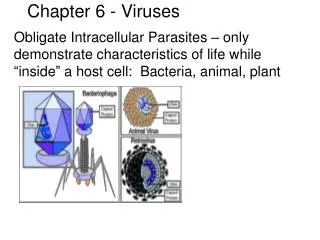
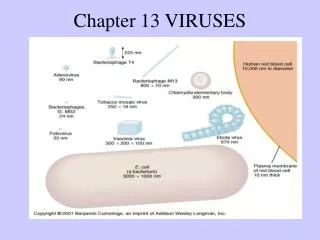
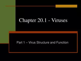
What are Viruses? • Obligate intracellular parasites • Viral components • Nucleic acids • Capsid • Envelope
Flu Attack How A Virus Invades Your Body http://www.youtube.com/watch?v=Rpj0emEGShQ&feature=related
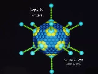
What are Viruses? • Obligate intracellular parasites • Viral components • Nucleic acids • Capsid • Envelope Enveloped icosahedral virus H and N of influenza, (such as, H5N1) Fig. 10.1 The components of an animal virus (a herpesvirus)
Virus Classification • 108 families so far • Pathogens for all life forms • Classification based upon • Nucleic acid type • Single strand vs. double strand chromosome • + versus – single strand • Enveloped versus “naked“ • RNA genomes occur only in viruses
Envelopes-enveloped viruses have a typical bilayer membrane outside of their capsids. The proteins of the membrane are typically derived from viral genes information whereas, the lipid is synthesized by the cellular machinery (i.e., there are no viral genes that code for the lipid moiety of the viral envelope). Envelopes often have glycoprotein projections termed spikes that are very important for attachment of the virus to a host cell receptor. Enveloped viruses are relatively sensitive to environmental insults such as drying, pH, freezing and thawing and many types of disinfectant , and are easily damaged whereas, non-enveloped viruses or naked viruses are often quite resistant to these insults. Hence, enveloped viruses are rarely spread via fomite (inanimate object) Fortunately HIV is an enveloped virus-Fortunate because that assures that it cannot survive very well outside the body. http://www.youtube.com/watch?v=NKoZfHLQu5M
Herpes simplex virus envelopment and release http://www.youtube.com/watch?v=bgj1YpevA6A&feature=related
Host range and specificity of viruses.-The host range of a virus refers to the spectrum of hosts that a virus can infect. Most viruses are limited to only one host and to only specific cells and/or tissues of that host. –(e.g., Polio)- exceptions, e.g, rabiesvirus Viral specificity is determined mainly by whether or not a virus can attach to a cell. Attachment depends on thepresence of specific receptor sites on the surfaces of host cells and on specific attachment structures on viral capsids (polio) or envelopes (H1N1; HIV)-flu, immunodefeciency virus. Specificity also depends on whether the host can supply the appropriate enzymes and other proteins the virus needs to replicate Additionally, virus specificity, refers to the specific kinds of cells a virus can infect, e.g, papillomaviruses, which cause warts, can only infect skin cells. Hepatoma virus, rabies virus, liver and nerve cells respectively. In contrast, cytomegalovirus (CMV) can cross placenta salivary glands, GI tract, liver, lungs and other organisms. CMV is an important virus that attacks the fetus.
Classification of Viruses. Currently the International Committee on Taxonomy of Viruses (ICTV) requires that the English common name, rather than a Latinized binomial term, be used to designate a viral species. For example, for rabies virus and HIV would be: family: rabies virus= Family: Rhabdoviridae; genus: Lyssavirus ; species: rabies virus HIV=Family: Retroviridae, genus: Lentivirus; species:human immunodeficiency virus (HIV).
Scientists seek to weaponize new family of bacteria to fight malaria By genetically modifying bacteria that they found to be uniquely associated with disease-carrying mosquitoes, scientists hope to create a new weapon to prevent transmission of malaria. The team isolated the bacterial strains from larvae of the mosquito Anopheles arabiensis, one of the most important spreaders of malaria in sub-Saharan Africa and surrounding areas. Thorsellia bacteria seem to be uniquely associated with disease-carrying mosquitoes When we discovered the first species of Thorsellia in a Kenyan malaria mosquito and decided to name the unique bacterium after Thorsell, we did not know that it would prove to be so common in mosquitoes." Since first discovering Thorsellia bacteria in Kenyan malaria mosquitoes, the scientists have also isolated strains from mosquitoes spreading malaria in Africa, Brazil, India and Iran, and in mosquitoes spreading West Nile virus in the US. It is unusual to find a new family of bacteria in this part of the family tree - it has only happened once before in the last 50 years. "We are looking for bacteria that live in the mosquito gut and which grow quickly when the mosquito has taken a blood meal. The idea is to genetically modify these bacteria to produce substances that stop malaria parasite development."
Scientists seek to weaponize new family of bacteria to fight malaria By genetically modifying bacteria that they found to be uniquely associated with disease-carrying mosquitoes, scientists hope to create a new weapon to prevent transmission of malaria. The team isolated the bacterial strains from larvae of the mosquito Anopheles arabiensis, one of the most important spreaders of malaria in sub-Saharan Africa and surrounding areas. Thorsellia bacteria seem to be uniquely associated with disease-carrying mosquitoes When we discovered the first species of Thorsellia in a Kenyan malaria mosquito and decided to name the unique bacterium after Thorsell, we did not know that it would prove to be so common in mosquitoes." Since first discovering Thorsellia bacteria in Kenyan malaria mosquitoes, the scientists have also isolated strains from mosquitoes spreading malaria in Africa, Brazil, India and Iran, and in mosquitoes spreading West Nile virus in the US. It is unusual to find a new family of bacteria in this part of the family tree - it has only happened once before in the last 50 years. "We are looking for bacteria that live in the mosquito gut and which grow quickly when the mosquito has taken a blood meal. The idea is to genetically modify these bacteria to produce substances that stop malaria parasite development."
Emerging Viruses Emerging Viruses- viruses that were previously endemic (low levels of infection in localized areas) or had “crossed species barriers”. 1. Polio virus- stable + stranded RNA virus-endemic with isolated cases. Growth of cities, crowding, pressure on the sanitary systems became a major health problem until vaccines were discovered. The outbreaks in the US were largely due to the superb sanitation system we developed. 2. HIV- likely came from crossover of SIV (simian immunodeficiency virus). 3. Hanta virus. Emerging problem in the southwest USA. ?----rodents---increased rodent population---feces--infection of humans 4. SARS (severe acute respiratory syndrome)- Coronavirus-that can cause severe upper respiratory disease and has a mortality rate of about 10% 5. Bird flu. H5N1- currently a major potential health problem 6. West Nile virus- It mainly infects birds, but is known to infect humans, horses, dogs, cats, bats, chipmunks, skunks, squirrels, and domestic rabbits. The main route of human infection is through the bite of an infected mosquito. 7. Might add Ebolavirus Based on recent history it is quite likely that the next emerging disease will be a virus.
Let us examine the strategy of replication of RNA viruses
Strategy of positive stranded RNA virus replication- both retrovirus and non-retrovirus Fig. 10.13 Replication of RNA viruses
negative How a negative stranded virus (rhabdovirus) replicates
Viral Replication General characteristics of replication 1. Adsorption 2. Penetration 3. Synthesis 4. Maturation 5. Release
Replication of bacteriophages d’Herelle and Twort given credit for identifying and naming bacteriophage (eaters of bacteria) Properties of bacteriophages 1. Either double-stranded or single-stranded RNA or DNA 2. Can be relatively simple or complex in structure 3. Can be used in antibacterial therapy because they are quite specific and can work against antibiotic resistant bacteria. Phage resistance can readily be overcome by selection. Russians routinely use phage therapy with very good results.
Bacteriophage Therapy – from Biotech Journal Bacteriophage therapy has many advantages over antibiotics. Bacteriophages are highly specific, where as antibiotics kill all bacteria without specificity, beneficial bacteria (e.g. in the intestinal tract) that perform crucial functions for the human body are also affected by antibiotics and harmful pathogens can then grow more easily. Secondary infections like the Pseudomonas species or Clostridium dificiledevelop in this way and cause severe diarrhea and colon infections. Bacteriophages can specifically target the harmful bacterium, eliminate it, and leave the beneficial bacteria intact. Bacteriophages cannot cause disease to humans, animals, or plants; they can only cause harm to bacteria. Furthermore, for almost all known bacterial species there exists one or more bacteriophages specific to that species. The bacteriophage therapy is currently being used to treat post-burn bacterial infections, which are a major problem for those recovering from the trauma of third-degree burns. Within 24 hours, burn patients can start suffering from opportunistic bacterial attacks. As an alternative to treating post-burn bacterial infections by antibiotics, bacteriophages have been in use in certain parts of the world, such as at Tbilisi in Georgia and in Poland, and this approach has now been more widely recognized. Results have shown that bacteriophage therapy has an 80% success rate against Enterococcus infections and up to 90% against Staphylococcus aureus, Pseudomonas aeruginosa, Escherichia coli and Klebsiellapneumoniae. Pseudomonas aeruginosa is the most common post-burn infection, and it is known to be notoriously resistant to a variety of antibiotics. For the most effective treatment of post-burn infections, a cocktail of bacteriophages is sprayed at the site of burns, this will reduce the chance of the bacteria developing resistance against the different bacteriophages. Bacteriophage solutions or aerosols can also be used to treat the surfaces and instruments in operating rooms as well as the skin of the surgical patient (prior to surgery). The above use not withstanding problems exist with the use of phage: 1. resistance 2. immunological response
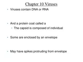
capsid with nucleic acid inside Tail sheath ATP-mediated contraction Fig. 10.10 Bacteriophages
Start http://www.youtube.com/watch?v=OxvqhneAX40&feature=related Fig. 10.11 Replication of a virulent bacteriophage
Bacteria exchange food via nanotubes A new study shows that some bacteria can form nanotubes between single cells that allow the cells to exchange essential nutrients or metabolites with each other. Now, writing in the journal Nature Communications, a team of scientists from several German research centers - including the Max Planck Institute for Chemical Ecology in Jena - reveals that bacteria exchange nutrients directly with each other through nanotubes strung between single cells. Their study investigates two species of bacteria: the gut microbe Escherichia coli, and the soil bacterium Acinetobacter baylyi. The scientists found that when cultured together, the bacteria were able to cross-feed each other - supplying to the other the amino acids that the other could not produce for itself.Then, they grew the two species of bacteria very close together but separated them with a filter so amino acids could not pass between them via the culture medium and there was no direct contact between the cells of the two species. In the second experiment, the bacteria died. The team concludes it showed that direct contact between cells is necessary for nutrient exchange and for both strains to thrive. When they looked at the culture containing the two species mixed together under an electron microscope, the researchers saw tiny filamentous nanotubes connecting individual cells. These were enabling the cells to exchange metabolites with each other. When the missing amino acid was introduced to the culture, the bacteria did not form nanotubes, suggesting that they only do so when they are "hungry" for the required nutrient, explains Kost. FOLLOWING SLIDE SHOWS THE NANOTUBES IN E. COLI
Replication of a virulent DNA bacteriophage 1. Adsorption- specific proteins in the phage tail fibersbind to specific receptor sites on the host cells. The fibers bend and allow the pins to touch the cell surface. 2. Penetration- The enzyme lysozyme (characterized by Sir Alexander Fleming), weakens the bacterial cell wall. And allows the viral DNA to be “injected” into the cytoplasms (either directly or into the periplasmic space and then into the cytoplasm). 3. Synthesis- once the phage DNA enters the cell the phage genes take control of the host cell’s metabolic machinery. Phage DNA is transcribed to mRNA, using the host cell’s machinery. The mRNA translated on host ribosomes, then directs the synthesis of capsid proteins and viral enzymes such as DNA polymerase that replicates the phage DNA. 4. Maturation - the parts of the phage are put together in a certain order but essentially it is a rapid process of assembly 5. Release- the enzyme lysozyme, which is coded for by a phage gene, breaks down the cell wall allowing viruses to escape. In the process
The time from adsorption to release is called the burst time; it varies from 20 to 40 minutes depending on the phage. The number of new virions released from each bacterial host represents the viral yield, or burst size.
The eclipse period represents the time after penetration through the biosynthesis of mature phages. The latent period represents the time after penetration through the release of mature phages. The number of viruses per infected cell is the viral yield or burst size. Fig. 10.12 Growth curve for a bacteriophage
The number of bacteriophages in a sample is assayed by spreading the sample out over a lawn of solid bacterial growth. When the phages replicate and destroy the bacterial cells, they leave a clear spot, called a plaque, in the lawn. The number of plaques corresponds roughly to the number of phages that were initially present in the sample. Fig. 10.13 Plaque assay
http://www.youtube.com/watch?v=_J9-xKitsd0&NR=1 lysogeny is very important for toxin production since in many instances the phage carries the toxin gene Fig. 10.15 Replication of a temperate bacteriophage
Replication of a temperate (lysogenic) bacteriophage Insertion of a lambda phage into a bacterium alters the genetic characteristics of the bacterium. Two genes present in the prophage produce proteins that repress virus replication. The prophage also containsanother gene that provides “immunity to infection by another phage. This process called lysogenic conversion, prevents the adsorption or biosynthesis of phages of the types whose DNA is already carried by the lysogen and Lysogenic conversion can be of medical significance because the toxic effects of some bacterial infections are caused by the prophages they contain, e.g., Corynebacterium diphtheriae and Clostridium botulinum which containprophage that code for their respective toxins.
Stages of animal virus infection with a DNA virus Adsorption penetration Synthesis Maturation Release DNA Animal Viruses • Chromosome replication in host cell nucleus • Cytoplasmic ribosomes for viral mRNA translation • Viral proteins must return to nucleus for maturation phase • Early vs. late transcription How do viruses avoid digestion by lysosomes?
Use RNA-dependent RNA polymerase • Cytoplasmic chromosomal replication and protein synthesis • + strands always needed for mRNA Synthesis. In picronavirus the (+) strand acts as mRNA. HIV budding from T-4 lymphocyte Fig. 10.17a Replication of RNA viruses-Polio
Use RNA-dependent DNA polymerase (Reverse transcriptase) • + strand chromosome acts as template for ds DNA provirus • Later transcription of provirus allows for virus production Synthesis. In the retroviruses, such as HIV, the two copies of (P) sense RNA do not act as mRNA but rather they are transcribed into ssDNA with the help of reverse transcriptase. HIV budding from T-4 lymphocyte Fig. 10.17b Replication of RNA viruses-HIV
Adsorption Figure 10.16 Viral recognition of an animal host cell a) Rhinoviruses have “canyons,: or depressions in the capsid that attach to specific membrane proteins on the host cell membrane b) HIV has specific envelope spikes (viral glycoproteins) that attach to a membrane protein receptor on the surface of specific host immune defense cells. http://student.ccbcmd.edu/courses/bio141/lecguide/unit3/viruses/adsorp_ev_fl.html
Rhinovirus (non-envelope) type attachment HIV (envelope) type attachment Fig. 10.18 Viral recognition of an animal host cell