Download
1 / 34
340 likes | 458 Views
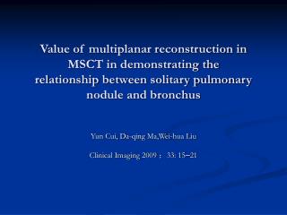
Value of multiplanar reconstruction in MSCT in demonstrating the relationship between solitary pulmonary nodule and bronchus. Yun Cui, Da-qing Ma,Wei-hua Liu Clinical Imaging 2009 ; 33: 15 – 21. Vocabulary. Nodule n. 1. 小瘤 ; 小结 2.( 藻 ) 节 ;( 菌 ) 瘤 3. 结核 Bronchus n. 支气管

E N D
Value of multiplanar reconstruction in MSCT in demonstrating therelationship between solitary pulmonary nodule and bronchus Yun Cui, Da-qing Ma,Wei-hua Liu Clinical Imaging 2009 ;33: 15–21
Vocabulary • Nodule n. 1.小瘤;小结2.(藻)节;(菌)瘤3.结核 • Bronchus n. 支气管 • Order 等级,阶层; 顺序,次序 • Bronchi 英['brɑnkai]美['brɑnkaɪ] (bronchus的复数)支气管 bronchial 英['brɔŋkiəl]美['brɑŋkɪəl] a. 支气管的 • Pneumonia 英[nju:'məunjə]美[nju'monjə] • abscess 英['æbsis]美['æbsɪs] n. 脓疮 • Hemangioma 英[hi:,mændʒi:'əumə]美[hi,mændʒɪ'omə] n.血管瘤 • Aspergilloma 曲霉肿
Lumen n. 1. 【物】流明2. 【解】内腔 • Obliterate 英 [ə'blitəreit]美 [ə'blɪtə,ret] • vt. 1. 擦掉...的痕迹;冲刷;消灭 2. 忘掉,忘却 • dislocate 英 ['disləkeit]美 ['dɪslə,ket] • vt. 1. 使移动位置;使脱臼 2. 弄乱 • Dilation n. 扩张;扩大部分 • Converge 英 [kən'və:dʒ]美 [kən'vɝdʒ] vi. 1. 会合;趋于会合[(+on/toward)] 2. 聚集;趋于同样结果 [(+on)] 3. 【数】收敛 vt. 1. 使向一点会合;使聚集
sequestration 英[sikwe'streiʃən]美[sɪkwɛ'streʃən] n. 1. 扣押,没收,接收 2. 隔离 Distomiasis 双盘吸虫病 • notch 英 [nɔtʃ]美[nɑtʃ] n. 1. 刻痕;槽口,凹口 2. 等,级 3.峡谷,山峡 • Spinous 英 ['spainəs]美 ['spaɪnəs]a.刺状的 • Taper 英 [‘teipə]美 [’tepɚ] n. 1. 逐渐变得尖细 2. 逐渐减少;逐渐变弱 3. 锥形物;尖塔[C] • Bifurcation 英 [,baifə:'keiʃən]美 [,baɪfɚ'keʃən] n. 分叉;分枝;分歧 • Bifurcate 英 ['baifə:keit]美 ['baɪfɚ,ket] vt. 1. 使分枝;使分叉 vi. 分枝;分叉 a. 分叉的
Introduction • To our knowledge, At present, with the application of thin-section scanning using multislice spiral computed tomography (MSCT), the lower-order bronchi can be observed clearly. • Furthermore, multiplanar recon-struction (MPR) of MSCT postprocessing enables the bronchi that can not be shown on axial images to be demonstrated continuously and wholly from various angles and increases visualization of the SPN–bronchus relation- ship (Fig.1).
Fig.1(A) Axial image showing a nodule in the left upper lobe,but no nodule–bronchus relationship is visualized.(B) Same case. A bronchus leading to the nodule is shown after the performance of MPR (arrow).
The purpose of our study was to investigate the value of MPR in MSCT in demonstrating the SPN–bronchus relationship for differentiating malignant from benign SPNs.
Materials and methods • We collected 148 SPN cases confirmed by operation, bronchoscopy, drug treatment, and follow-up between September 2006 and September 2007. • The patients (103 males and 45 females) ranged in age from 18 to 80 years (average, 56 years). • The greatest diameter of the nodules, as measured on CT scans, ranged from 1.1 to 4.0 cm (average, 2.6 cm).
3.1 Bronchi shown on axial and MPR images • The SPN–bronchus relationship was positive in 43 of 148 cases (29%) on axial images, and in up to 95 cases (64%) on MPR images. • It suggests that MPR can elevate the visualization of bronchi and can show the bronchial stereo-structure continuously and wholly from various angles. • The 95 cases included 62 malignant nodules and 33 benign nodules.
Malignant nodules consisted of adenocarcinoma (AC; n=33), squamous carcinoma (SC; n=16), bronchioal-veolar carcinoma (BAC; n=3), carcinosarcoma (n=2), adenosquamous carcinoma (AdCa; n=1), small-cell carcinoma (SCC;n=1; Fig.2), and metastatic tumor (MT; n=6). • Benign nodules consisted of tuberculoma (n=14), chronic pneumonia (n=6), globular pneumonia (n=5), lung abscess (n=2), sclerosing hemangioma (n=2), inflammation pseudonoma (n=1), aspergilloma (n=1), pulmonary sequestration (n=1), and pulmonary distomiasis (n=1).
With this CT protocol, the bronchi related to the nodules of the 95 cases were mainly of fourth to sixth orders. • There was no significant difference between malignant and benign nodules.
Fig.2SCC in the right upper lobe.The bronchus is obstructed by the tumor(arrow).
3.2 Nodule size and occurrence of the SPN–bronchus relationship • The results of our study showed that the bronchi were visualized in all nodules greater than 3 cm, in 87% of nodules less than 3 cm and greater than 2 cm, and in 37%of nodules less than 2 cm ( Table 1). • It indicates that nodule size is a significant factor for determining whether it has the bronchus sign or not.
Fig.3 Tuberculoma in the right upper lobe. A bronchus leading to the tuberculoma is seen (arrow).
Fig.4Tuberculoma in the right upper lobe.The twisted bronchus with a thickened wall (arrows).
Fig.5Moderately differentiated AC in the left lower lobe. At the site of the bronchus entering the nodule, the tumor is associated with the notch (arrow).
Fig.6Poorly differentiated AC in the left upper lobe.The bronchus is connected with the spinous process of the tumor (arrow).
3.3 Morphologic characteristics of the SPN–bronchus relationship • We classified the SPN–bronchus relationship into four types based on the classification made by Tsuboi and Choi etal: Type I (bronchus cut off at the edge of SPN), Type II (bronchus cut off in the SPN) (Table 2), Type III (bronchus running through the whole SPN), Type IV (bronchus running at the periphery of the SPN). • Each type was then classified into several subtypes. • Many SPNs showed more than one type of SPN–bronchus relationship. • No significant difference was observed between malignant and benign nodules in each type.
3.4 SPN–bronchus relationship and type of lung cancer • AC showed all four types of SPN–bronchus relationship. • SC showed Type I,Type II,and Type IV patterns,but no Type III pattern. • There was no significant difference between SC and AC in Type I, Type II,and Type IV patterns. • Type III pattern was more common in AC but was not observed in SC ( P< .05). • MT (n=6) most often had a Type I pattern (5 of 6) (Table 3).
3.5 SPN–bronchus relationship and degree of differentiation of lung cancer • The degrees of differentiation of 38 AC and SC were confirmed (Table 4). • We analyzed the appearance of the bronchus and found that the bronchus'connection with the spinous process of the SPN was more frequently seen in poorly differentiated lung cancer than in moderately differentiated lung cancer, and that Type III pattern was more common in moderately differentiated lung cancer than in poorly differentiated lung cancer ( P< .05). • No statistical significance was shown between moderately and poorly differentiated lung cancers in other patterns.
In malignant SPN, tumor cells proliferate and invade the surrounding lung parenchyma continuously ( Fig.7). • As a result, the adjacent bronchi are commonly obstructed with tapered narrowing or flat ends ( Fig.8). • A bronchus obstructed abruptly can also occur in benign SPNs, especially in tuberculoma, and there is often a capsule around the tuberculoma that can be confluent with the bronchus. • Different parts of a malignant nodule usually grow at different speeds. • Therefore, at the site of a bronchus entering the nodule, the nodule can associate with the notch because of a speed relatively slower growing than those in other sites due to blockage by the bronchus.
In addition, at the tumor–bronchus interface, the nodule can also associate with the spinous process due to the tumor cells growing along with peribronchial tissue and having a faster-growing speed. • Besides, in benign SPN, especially in tuberculoma, caseous content can be discharged through the bronchus to thicken the bronchial wall, which may be associated with the bronchus being twisted . • Benign SPN cannot invade the parenchyma, so the bronchus in or around the nodules is usually patent (Fig.9) or compressed,with an intact and regular wall.
However, in malignant SPN, the tumor cells always infiltrate the mucosa or submucosa of the bronchus, resulting in a thickened wall and an irregularly narrowed lumen (Figs.10–14). • In lung cancer, there is often fibrous degeneration, resulting in the bronchial lumen being dilated and in the bronchus converging with it (Fig.15).
Fig.8Moderately differentiated AC in the left lobe.The bronchus is cut off with a tapered narrowing after entering the tumor (arrow).
Fig.9Inflammation pseudonoma in the right lower lobe.The bronchus in the nodule runs naturally with a regular wall and lumen (arrow).
Fig.10Moderately differentiated AC in the right upper lobe.The bronchi in the tumor run rigidly (arrow).
Fig.11Moderately differentiated AC in the right lower lobe.The bronchus in the tumor is irregularly narrowed (arrow)
Fig.12Moderately differentiated AC in the right middle lobe.The bronchial lumen beside the tumor is irregularly narrowed (arrow)
Fig.14Moderately differentiated SC in the left upper lobe.The bronchus beside the tumor is invaded with an obliterated lumen (arrow).
Fig.15Moderately differentiated AC in the right upper lobe.The bronchi converging at the tumor are seen (arrows).
conclusion • MPR can increase the demonstration of the SPN–bronchus relationship. • The SPN–bronchus relation-ship is valuable in determining the nature and the degree of differentiation of SPN.