Asthma
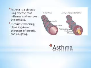
Asthma. Dr. Tara Husain. Definition;. is defined as reversible obstruction of large and small airways as a result of hyper-responsiveness to various immunologic and non immunologic stimuli. Risk factors. Environment; inhaled allergens respiratory viral infections

Asthma
E N D
Presentation Transcript
Asthma Dr. Tara Husain
Definition; • is defined as reversible obstruction of large and small airways as a result of hyper-responsiveness to various immunologic and non immunologic stimuli
Risk factors • Environment; • inhaled allergens • respiratory viral infections • chemical and biologic air pollutants • Biological and genetic factors
EARLY CHILDHOOD RISK FACTORS FOR PERSISTENT ASTHMA • Parental Asthma • History of other allergy • Sever lower respiratory tract infection • Wheezing apart from colds • Male gender • Low birth weight • Exposure to tobacco smoke • Reduced lung function at birth
Infants are more susceptible to airway obstruction due to; • Smaller airway size. • Lower elastic recoil of the lung. • Decreased smooth muscle support of the small airways. • Relative mucus gland hyperplasia. • Decreased collateral channels of ventilation between the alveoli
Asthma triggers; • viral infections • Aeroallergens (animal dander, dust mites) • Seasonal aeroallergens ( Pollens) • tobacco smoke • Air pollutants;( Ozone, Sulfur dioxide) • Strong or noxious odors • Cold , dry air • Exercise • Crying, laughter, hyperventilation
Aggravating factors in asthma • Gastroesophageal reflux • Rhinitis • Sinusitis
Clinical Manifestations • Intermittent dry coughing and expiratory wheezing are the most common symptoms • shortness of breath and chest tightness • symptoms can be worse at night • Daytime symptoms, often linked with physical activities • symptoms can be subtle ; (limitation of physical activities, general fatigue ,or sleep disturbance) • Lack of improvement with bronchodilator and corticosteroid is inconsistent with the diagnosis of asthma
Examination; • expiratory wheezing and a prolonged expiratory phase • Decreased breath sounds in some of the lung fields, commonly the right lower posterior lobe, are consistent with regional hypoventilation owing to airways obstruction. • Crackles and rhonchi can sometimes be heard, resulting from excess mucus production and inflammatory exudate in the airways
Differential diagnosis; • Chronic rhinitis • Sinusitis • Adenoidal or tonsillar hypertrophy • Laryngotracheobronchomalacia • Laryngotracheobronchitis • Vocal cord dysfunction • Foreign body aspiration • Viral bronchiolitis • Gastroesophageal reflux • Tuberculosis • Heart failure
Diagnosis; • Mainly clinical • Pulmonary function test • Radiography • Skin test • Radioallergosorbent (RASTA) test
Pulmonary function test Tidal volume • Is the lung volume representing the normal amount of air displaced between normal inhalation and exhalation with no extra effort applied
Total lung capacity; • is the maximum volume to which the lung can be expanded with the greatest possible inspiratory effort; • TLC= VC +RV
Vital capacity; • Is the maximum amount of air the person can expel from the lung after first filling the lung to the maximum extent
Functional residual volume and residual volume; • Functional residual capacity; is the volume of air present in lung at the end of passive expiration • Residual volume; is the amount of air left in the lung after maximum exhalation
Spirometry • objective measure of airflow limitation • Measures the forced expiratory volume in one second. • usually feasible in children > 6 yr • FEV1 is within 5% on 3 attempts, then the highest FEV1 effort of the 3 is used • Normal values are based on height, gender, and ethnicity. • typically decreases in asthmatics by > 15% after exercise challenge
Pulmonary function test; • useful in evaluating an older child’s functional status, although they rarely yield an etiological diagnosis • Abnormalities described in form of obstructive or restrictive lung disease
obstructive disease; there is low flow rate and increased residual volume RV and functional residual volume FRV • Restrictive disease; there is low vital capacity and total lung capacity with relative preservation of flow rates and FRV. • A significant increase in pulmonary function test after bronchodilator therapy indicate reactive airway disease
Chest X-ray; • often appear to be normal, aside of subtle nonspecific finding of hyperinflation and peribronchial thickening. • CXR is not routine in asthmatic patients
Indications of chest x-ray; 1- First attack of bronchospasm. 2- localized wheeze or other finding on osculation. 3- High grade fever or toxic appearance. 4- Sudden deterioration in patients condition despite treatment with signs suspecting pneumothorax. 5- suspicion of foreign body.
Skin test; • There are 2 methods; 1-Prick or puncture technique. 2-Intradermal injection technique
Radioallergosorbent (RASTA) test; • This is an in vitro test to determine the presence of antigen specific IgE in serum of the patient.
asthma masqueraders • aspiration pneumonitis • bronchiolitis obliterans • cystic fibrosis • allergic bronchopulmonary mycoses (aspergillosis) • ciliary dyskinesias • immune deficiencies