Download
1 / 26
350 likes | 1.04k Views
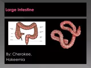
Anatomy of the Large Intestine. The large intestine ( colon ) extends from the ileocecal sphincter to the anus. Its subdivisions include the cecum, colon, rectum , and anal canal . Hanging inferior to the cecum is the appendix . Inflammation of the appendix is called appendicitis .

E N D
Anatomy of the Large Intestine • The large intestine (colon) extends from the ileocecal sphincter to the anus. • Its subdivisions include the cecum, colon, rectum, and anal canal. • Hanging inferior to the cecum is the appendix. • Inflammation of the appendix is called appendicitis. • A ruptured appendix can result in gangrene or peritonitis, which can be life-threatening conditions.
Anatomy of Large Intestine • 5 feet long by 2½ inches in diameter • Ascending & descending colon are retroperitoneal • Rectum = last 8 inches of GI tract anterior to the sacrum & coccyx • Anal canal = last 1 inch of GI tract • internal sphincter----smooth muscle & involuntary • external sphincter----skeletal muscle & voluntary control
Mechanical Digestion in Large Intestine • Mechanical movements of the large intestine include haustral churning, peristalsis, and mass peristalsis. • Peristaltic waves (3 to 12 contractions/minute) • haustral churning----relaxed pouches are filled from below by muscular contractions (elevator) • gastroilial reflex = when stomach is full, gastrin hormone relaxes ileocecal sphincter so small intestine will empty and make room • gastrocolic reflex = when stomach fills, a strong peristaltic wave moves contents of transverse colon into rectum by Mass peristalsis
Chemical Digestion in Large Intestine • No enzymes are secreted only mucous • Bacteria ferment • undigested carbohydrates into carbon dioxide & methane gas • undigested proteins into simpler substances (indoles)----odor • turn bilirubin into simpler substances that produce color • Bacteria produce vitamin K and B in colon • Converts chyme into feces
Functions of the Large intestinal Mucosa • Goblet cells: create mucus that lubricates colon and protects mucosa. • Absortive cells: Maintains water balance, solidifies feces, absorbs vitamins and some ions
Absorption & Feces Formation in the Large Intestine • Some electrolytes---Na+ and Cl- • After 3 to 10 hours, 90% of H2O has been removed from chyme • Feces are semisolid by time reaches transverse colon • Feces = dead epithelial cells, undigested food such as cellulose, bacteria (live & dead)
Absorption and Feces Formation in the Large Intestine • The large intestine absorbs water, electrolytes, and some vitamins. • Feces consist of water, inorganic salts, sloughed-off epithelial cells, bacteria, products of bacterial decomposition, and undigested parts of food. • Although most water absorption occurs in the small intestine, the large intestine absorbs enough to make it an important organ in maintaining the body’s water balance.
Defecation Reflex • The elimination of feces from the rectum is called defecation. • Defecation is a reflex action aided by voluntary contractions of the diaphragm and abdominal muscles. The external anal sphincter can be voluntarily controlled (except in infants) to allow or postpone defecation.
Defecation • Gastrocolic reflex moves feces into rectum • Stretch receptors signal sacral spinal cord • Parasympathetic nerves contract muscles of rectum & relax internal anal sphincter • External sphincter is voluntarily controlled
Defecation Problems • Diarrhea = chyme passes too quickly through intestine • H20 not reabsorbed • Constipation--decreased intestinal motility • too much water is reabsorbed • remedy = fiber, exercise and water • Clinical Concerns • Colonoscoy is the visual examination of the lining of the colon using an elongated, flexible, fiberoptic endoscope. • Occult blood test is to screen for colorectal cancer.
PANCREAS • The pancreas is divided into a head, body, and tail and is connected to the duodenum via the pancreatic duct (duct of Wirsung) and accessory duct (duct of Santorini). • Pancreatic islets (islets of Langerhans) secrete hormones and acini secrete a mixture of fluid and digestive enzymes called pancreatic juice.
Accessory organs of the GI Tract Pancreas: Produces 1.2L to 1.5L of pancreatic juices daily. Pancreatic juice consists of a bicarbonate solution containing salts and digestive enzymes. Bicarbonate helps buffer acidic chyme from the stomach
Histology of the Pancreas • Acinar cells: Secrete pancreatic juice, a mixture of bicarbonate fluid and digestive enzymes. • Islet of Langerhans: Alpha cells- glucagon Beta cells- insulin Delta cells- somatostatin F-cells- pancreatic polypeptide Acini Islet of Langerhans
Neural and Hormonal Control of the Pancreas • Secretin: • acidity in intestine causes increased sodium bicarbonate release • GIP: • fatty acids & sugar causes increased insulin release • CCK: • fats and proteins cause increased digestive enzyme release
LIVER AND GALLBLADDER • The liver is the heaviest gland in the body and the second largest organ in the body after the skin. • Anatomy of the Liver and Gallbladder • The liveris divisible into left and right lobes, separated by the falciform ligament. Associated with the right lobe are the caudate and quadrate lobes. • The gallbladderis a sac located in a depression on the posterior surface of the liver.
Histology of the Liver • The lobes of the liver are made up of lobules that contain hepatic cells (liver cells or hepatocytes), sinusoids, stellate reticuloendothelial (Kupffer’s) cells, and a central vein. • Bile is secreted by hepatocytes. • Bile passes into bile canaliculi to bile ducts to the right and left hepatic ducts which unite to form the common hepatic duct. • Common hepatic duct joins the cystic duct to form the common bile duct which enters the hepatopancreatic ampulla.
Pathway of Bile Secretion • Bile capillaries • Hepatic ducts connect to form common hepatic duct • Cystic duct from gallbladder & common hepatic duct join to form common bile duct • Common bile duct & pancreatic duct empty into duodenum
Accessory organs of the GI Tract Liver: Produces .8L to 1.0L of bile per day • yellow-green in color & pH 7.6 to 8.6 • Components • water & cholesterol • bile salts = Na & K salts of bile acids • bile pigments (bilirubin) from hemoglobin molecule • globin = a reuseable protein • heme = broken down into iron and bilirubin
Bile - Overview • Hepatic cells (hepatocytes) produce bile that is transported by a duct system to the gallbladder for concentration and temporary storage. • Bile is partially an excretory product (containing components of worn-out red blood cells) and partially a digestive secretion. • Bile’s contribution to digestion is the emulsification of triglycerides. • The fusion of individual crystals of cholesterol is the beginning of 95% of all gallstones. Gallstones can cause obstruction to the outflow of bile in any portion of the duct system. Treatment of gallstones consists of using gallstone-dissolving drugs, lithotripsy, or surgery.
Bile - Overview • The liver also functions in carbohydrate, lipid, and protein metabolism; removal of drugs and hormones from the blood; excretion of bilirubin; synthesis of bile salts; storage of vitamins and minerals; phagocytosis; and activation of vitamin D. • In a liver biopsy a sample of living liver tissue is removed to diagnose a number of disorders.
Major Functions of the liver • Carbohydrate metabolism: maintains blood sugar levels. a. Low Sugars levels: (control- glucagon) glycogenolysis glycogen > glucose b. High sugars levels: (control- insulin) glycogenesis glucose > glycogen • Lipid metabolism a. Produce fats: lipogenesis b. Break down fats: lipolysis, beta oxidation c. Synthesize cholesterol d. Stores triglycerides
Major Functions of the Liver • Protein metabolism: a. Synthesize most plasma proteins such as clotting proteins b. Deaminate amino acid: remove NH2 • Processes drugs, hormones, and alcohol • Excretes bilirubin (derived from the heme unit of recycled red blood cells) • Storage of Vitamins (A, B12, D, E, and K) and iron • Phagocytosis of aged red and white blood cells and some bacteria by Kupffer’s (reticuloendothelial) cells • Activation of Vitamin D • Stores iron and copper