Download

1 / 25
270 likes | 809 Views
Large intestine. Ileocaecal junction. Indigestible food residues from the ileum are propelled by peristalsis into the distended first part of the large intestine, the caecum , through the cone-shaped ileocaecal valve.

E N D
Ileocaecal junction • Indigestible food residues from the ileum are propelled by peristalsis into the distended first part of the large intestine, the caecum, through the cone-shaped ileocaecal valve. • There is an abrupt transition in the lining of the valve from the small intestinal villiform pattern S to the glandular form in the large intestine L.
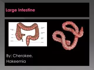
Large Intestine • The large intestine or bowel consists of a mucosal membrane with no folds except in its distal (rectal) portion and no villi • The mucosa is penetrated throughout its area by tubular intestinal glands lined by goblet and absorptive cells, with a small number of enteroendocrine cells
The absorptive cells or colonocytes are columnar and have short, irregular microvilli • Stem cells for the epithelium of the large bowel are located in the bottom third of each gland. • The large intestine is well suited to its main functions: absorption of water, formation of the fecal mass from undigestible material, and production of mucus that lubricates the intestinal surface.
The tall columnar absorptive cells have oval basal nuclei; in contrast, goblet cell nuclei are small and condensed. • Stem cells at the base of the glands continually replace the epithelium.
The mucosa has shallow plicae but no villi. • The muscularis has two layers, but the outer longitudinal layer consists only of three distinct bundles of muscle fibers called taeniae coli (ribbons of the colon). • These bands cause the colon wall to form a series of sacs called haustra. • The serosa of the colon is continuous with that of the supporting mesenteries and displays a series of suspended masses of adipose tissue called omental appendages.
Lamina propria fills the space between the glands and contains numerous blood and lymphatic vessels into which water is absorbed. • The lamina propria also contains collagen as well as lymphocytes and plasma cells. • These form part of the defence mechanisms against invading pathogens along with intraepithelial lymphocytes and the lymphoid aggregates LA, which are smaller than Peyer's patches, found in the lamina propria and submucosa
Rectum • At the distal end of the rectum, the anal canal, the mucosa and submucosa are highly vascularized, with venous sinuses, and are folded as a series of longitudinal anal folds with intervening anal sinuses. • Fecal material accumulates in the rectum is eliminated by muscular contraction, including action of an internal anal sphincter of smooth (involuntary) muscle and an external sphincter of striated (voluntary) muscle.
The mucosa is the same from caecum to rectum. It is folded in the non-distended state but it does not exhibit distinct plicaecirculares like those of the small intestine. • Immediately above the anal valves, the mucosa forms longitudinal folds called the columns of Morgagni.
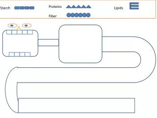
The large intestine is inhabited by a variety of commensal bacteria that further degrade food residues. Bacterial degradation is an important mechanism for the digestion of cellulose in ruminants, but in humans most cellulose is excreted. Small quantities of fat-soluble vitamins derived from bacterial activity are absorbed in the large intestine
The appendix is a small blind-ended tubular sac extending from the caecum just distal to the ileocaecal junction. • The mesenteries conduct blood vessels, lymphatics and nerves to and from the gastrointestinal tract. • The most characteristic feature of the appendix, particularly in the young, is the presence of masses of lymphoid tissue in the mucosa and submucosa.
The lamina propriaLP and upper submucosaSM are diffusely infiltrated with lymphocytes. • Note that the mucosal glands are much less closely packed than in the large intestine. • the lymphoid tissue also forms follicles F often containing germinal centres • These follicles bulge into the lumen and, like the follicles of Peyer's patches in the small intestine, are invested by a simple epithelium of M cells which presumably facilitates sampling of antigen in the lumen.
The rectum is the short dilated terminal portion of the large intestine. • The rectal mucosa RM is the same as the rest of the large bowel except that it has even more numerous goblet cells. • At the recto-anal junction J, it undergoes an abrupt transition to become stratified squamous epithelium SS in the anal canal. • Branched tubular circumanal glands open at the recto-anal junction into small pits at the distal ends of the columns of Morgagni
The anal canal forms the last 2 or 3 cm of the gastrointestinal tract and is surrounded by voluntary muscle that forms the anal sphincter. • Here, the stratified squamous epithelium undergoes a gradual transition to skin containing sebaceous glands and large apocrine sweat glands
Anus • In the anal region, the mucous membrane forms a series of longitudinal folds, the anal columns • About two cm above the anal opening, at the recto-anal junction, the lining of the mucosa is replaced by stratified squamous epithelium • In this region, the lamina propria contains a plexus of large veins that, when excessively dilated and varicose, can produce hemorrhoids.