Download
1 / 18
240 likes | 483 Views
ANATOMY OF THE SMALL INTESTINE. Dr. Jamila El- Medany. OBJECTIVES. At the end of the lecture, students should: List the different parts of small intestine.

E N D
ANATOMY OF THE SMALL INTESTINE Dr. Jamila El-Medany
OBJECTIVES At the end of the lecture, students should: • List the different parts of small intestine. • Describe the anatomy of duodenum, jejunum & ileum regarding: the shape, length, site of beginning & termination, peritoneal covering, arterial supply & lymphatic drainage. • Differentiate between each part of duodenum regarding the length, level & relations. • Differentiate between the jejunum & ileum regarding the characteristic anatomical features of each of them.
What is MESENTERY? Anterior abdominal wall Loop of intestine Posterior abdominal wall
FREE (MOVABLE) PART (WITH MESENTERY) JEJUNUM & ILEUM FIXED (Retro peritoneal) PART (NO MESENTERY) DUODENUM 1 2
DUODENUM • SHAPE:C-shaped loop • LENGTH: 10 inches • BEGINNING: at pyloro-duodenal junction • TERMINATION:at duodeno-jejunal flexure • PERITONEAL COVERING: retroperitoneal
PARTS • The duodenum is divided into (4) parts: • 1st :Superior. • 2nd: Descending (vertical). • 3rd: Inferior (Horizontal) • 4th : Ascending
Structures Related pancreas psoas
RELATIONS OF FIRST PART 3) 2) 1) X X Anterior Liver Posterior 1)Bile duct 2) Gastroduodenalartery 3)Portal vein
RELATIONS OF SECOND PART Anterior 1)Liver 2)Transverse Colon 3)Small intestine Posterior Right kidney X Lateral R Colic Flexure Medial Pancreas
OPENINGS IN SECOND PART OF DUODENUM • Common opening of bile duct & main pancreatic duct: on summit of major duodenal papilla. • Opening of accessory pancreatic duct (one inch higher): on summit of minor duodenal papilla.
RELATIONS OF THIRD PART • Anterior: a)Small intestine b) Superior mesenteric vessels • Posterior: 1) Right psoas major 2) Inferior vena cava 3) Abdominal aorta 4) Inferior mesenteric vessels. 1 2 3
RELATIONS OF FOURTH PART • Anterior: Small intestine • Posterior: Left psoas major psoas
Blood Supply & Lymph drainage • Because the duodenum is derived from both: Foregut & Midgut, • It has its Arterial Supply from : • Celiac & Superior mesenteric arteries. • Venous Drainage to : • Superior mesenteric& Portal veins. • LYMPHATIC DRAINAGE: Celiac & Superior mesenteric lymph nodes.
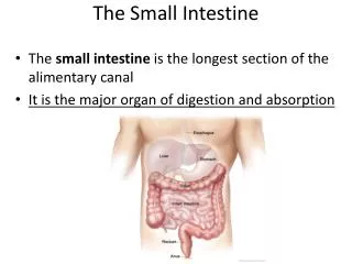
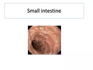
JEJUNUM & ILEUM • SHAPE:Coiled tube • LENGTH: 6 meters (20 feet) • BEGINNING: at Duodeno-jejunal flexure • TERMINATION:at Ilieo-caecal junction • EMBRYOLOGICAL ORIGIN: Midgut • Blood SUPPLY: Superior mesenteric A & V • LYMPHATIC DRAINAGE: Superior mesenteric lymph nodes