Download
1 / 29
290 likes | 429 Views
Learn about the structure and function of the large intestine, including mucosa, submucosa, muscularis externa, and serosa. Discover the histology of appendix and its variations compared to the large intestine.

E N D
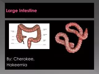
LARGE INTESTINE • Extends from the distal end of the ileum to the anus. • Consists of caecum, ascending colon, transverse colon, descending colon, sigmoid colon, rectum and anal canal. • Absorbs fluids and salts from the gut contents.
MUCOSA • Absence of villi • Raised into numerous cresentic mucosal folds. • Epithelium:- • Columnar cells with brush border. • Large number of Goblet cells. • Enteroendocrine cells/enterochromaffin cells/argentaffin cells. • Stem cells.
Tubular intestinal glands/Crypts of lieberkuhn in lamina propria also contain columnar cell, goblet cell, entero endocrine cell and stem cell. • The columnar cells with striated borderabsorb excess water and electrolytes. • The goblet cell number increases caudally down the intestine. • They secrete mucus that lubricates and facilitates the passage of the semisolid intestinal contents. • Stem cells differentiate and replace the damaged epithelial cells.
ENTEROENDOCRINE CELLS/ ENTEROCHROMAFFIN CELLS/ARGENTAFFIN CELLS: • The cells contain black infranuclear granules. • They stain with silver salts. • They produce amines like 5-HT. • These cells are positive to chromaffin reaction (with potassium dichromate) and hence called enterochromaffin cells.
SUB MUCOSA • Sub mucous layer: contains blood vessels, nerves, lymphatics, areolar connective tissue. Submucosa often contains fat cells with pas positive granules termed muciphages.
MUSCULARIS EXTERNA • Inner circular and outer longitudinal layer of muscle. • Outer longitudenal layer is unusual. • Most of the fibres form three thick bands called taenia coli. • As the taenia are shorter in length the intestine here is sacculated called haustrations. • A thin layer of longitudinal fibres is present in the intervals between the taenia coli.
SEROSA • The serous layer is the peritoneal covering. • It is missing over the posterior aspect of ascending and descending colon and present in other parts. • It has pouch like processes filled with fat called appendices epiploicae.
Narrowest part of the gut. • Has a narrow lumen and no villi seen. • The cryptsare poorly developed. • The longitudenal muscle coat is uniformly thick and has no taenia coli. • The lamina propria and submucosa is full of lymphoid nodules. • The lymphoid tissue is absent at birth and reaches a maximum at ten years and then progressively decreases with age.
MUCOSA Mucous membrane similar to large intestine: simple columnar with large number of goblet cells. Lamina propria consists of much more lymphatic tissue extending onto submucosa and has fewintestinal glands or Crypts of lieberkuhn. Muscularis mucosa is not well developed and missing in some areas.
SUB MUCOSA Consists of abundant lymphatic nodules, connective tissue, blood vessels and nerve plexus. MUSCULARIS EXTERNA Inner circular muscle fibers. Outer longitudinal muscle fibers and no taenia coli. SEROSA Made up of mesentery.
Mucosa Simple columnar epithelium with numerous goblet cells Lamina propria Crypts of Lieburkuhn Confluent lymphatic nodules
Summary: • Proximal to distal 1. Increase in lymphocytes. 2. Increase in Goblet cells. 3. Decrease in villi.