Download
1 / 19
190 likes | 344 Views
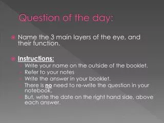
Question of the day:. Name the 3 main layers of the eye, and their function. Instructions: Write your name on the outside of the booklet. Refer to your notes Write the answer in your booklet. There is no need to re-write the question in your notebook.

E N D
Question of the day: • Name the 3 main layers of the eye, and their function. • Instructions: • Write your name on the outside of the booklet. • Refer to your notes • Write the answer in your booklet. • There is no need to re-write the question in your notebook. • But, write the date on the right hand side, above each answer.
Rays from the top of the candle enter the eye through thePUPIL opening and then hit theLENS. Once they hit the lens the rays are bent in such a way that they end upIN FOCUS directly on theRETINA The Formation of an Image on the Retina: Light rays always travel in a STRAIGHT LINE.
The same happens to the light rays from the bottom of the candle. • What do you notice about the image on the retina? • It isUPSIDE DOWN!! • All of the rods and cones located on theRETINAare then stimulated and send that message through the optic nerve to our brain. • The brains then flips the image around so we see the candle right side up.
The final image: Specialized receptors and BRAIN AREAS process different COMPONENTS of vision. THEN the brainPUTS ALL THE INFORMATION BACK TOGETHER
The Eye and the Image: Steps in seeing • The Cornea Light enters the eye through the cornea (the transparent front part of the eye that covers the iris and pupil). The cornea actually contributes most of the eye'sFOCUSING POWER, BUT its focus is fixed.
2. The Iris and pupil Controlling light levels: Your eyes are verySENSITIVEand can beDAMAGEDby harsh light. TheIRISis aSMOOTH MUSCLE (sphincter) responsible for controlling how muchLIGHTenters the eye. ThePUPILis the opening left by the iris (not really a “part”)
In aDARKroom where there is not much light the pupil will becomeLARGEto allow a lot of light in. On a very sunny day when there is a lot of light the pupil will becomeSMALLERto allowLESSlight in.
If the pupil is a hole that lets light into the eye, why does it appear black? It appears black because light rays entering the pupil are absorbed by tissues inside the eye. The pupil dilates (gets wider) in response to extreme emotional situations such as fear or pain. The same thing happens in response to loads on working memory and attention. Such stimuli awakens your “fight or flight” response of your CNS. Brain chemicals (neurotransmitters) such as adrenaline cause immediate physical reactions.
3. The Lens (Changing lens thickness) • CILIARYmuscles are attached to the lens, whenCONTRACTEDthey pull the lens thin
Far Objects: • When looking at objectsFAR AWAY the CILIARYmuscles contract and the lens becomes THIN, this way the rays appear on the retina and the image is clear. Close Objects: • When objects are CLOSEtheCILIARYmusclesRELAX and make the lens FATallowing the rays to project on the retina and the image is clear.
Why do we need glasses? Our RETINAis like the screen of an overhead projector. If the image does notLANDon the screen properly it will lookBLURRED. Light rays will bend and landSHARPLYon the retina (screen) only if theRETINAis the proper DISTANCEfrom the lens.
The eyeball is tooSHORTand the rays are projectedBEHINDthe retina so vision is blurred. This can be corrected by placing a CONVEX lens (glasses) in front of the eye. If you are Far-Sighted (hypermetropia)
The eyeball is too long and the image appears IN FRONTof the retina so vision is blurred. This can be corrected by placing a CONCAVElens (glasses) in front of the eye. If you are Near-Sighted (myopia)
Myopia Hyperopia Normal View Near Sighted Far Sighted
Convex lens: pre-bends light INWARD to shorten the focal point Concave lens: pre-bends light OUTWARD to lengthen the focal point
Seeing Color: • The type of photoreceptors responsible for seeing color are theCONES. • There are3types of cones, each with a heightened sensitivity to eitherRED, GREEN,orBLUE. • Every colour we see is some combination of these three colors. • Colorblindness is the inability to distinguish between certain colors and is caused by a chemical disorder in the cones.