Download
1 / 8
80 likes | 135 Views
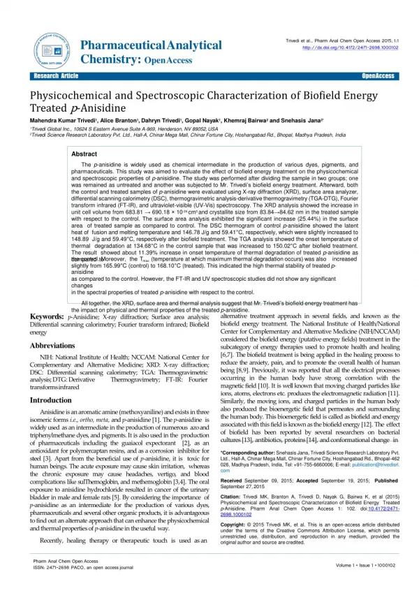
The p-anisidine is widely used as chemical intermediate in the production of various dyes, pigments, and pharmaceuticals. This study was aimed to evaluate the effect of biofield energy treatment on the physicochemical and spectroscopic properties of p-anisidine. The study was performed after dividing the sample in two groups; one was remained as untreated and another was subjected to Mr. Trivedi’s biofield energy treatment. Afterward, both the control and treated samples of p-anisidine were evaluated using X-ray diffraction (XRD), surface area analyzer, differential scanning calorimetry (DSC), thermogravimetric analysis-derivative thermogravimetry (TGA-DTG), Fourier transform infrared (FT-IR), and ultraviolet-visible (UV-Vis) spectroscopy. The XRD analysis showed the increase in unit cell volume from 683.81 → 690.18 × 10⁻²⁴ cm3 and crystallite size from 83.84→84.62 nm in the treated sample with respect to the control. The surface area analysis exhibited the significant increase (25.44%) in the surface area of treated sample as compared to control. The DSC thermogram of control p-anisidine showed the latent heat of fusion and melting temperature and 146.78 J/g and 59.41°C, respectively, which were slightly increased to 148.89 J/g and 59.49°C, respectively after biofield treatment. The TGA analysis showed the onset temperature of thermal degradation at 134.68°C in the control sample that was increased to 150.02°C after biofield treatment. The result showed about 11.39% increase in onset temperature of thermal degradation of treated p-anisidine as compared to the control. Moreover, the Tmax (temperature at which maximum thermal degradation occurs) was also increased slightly from 165.99°C (control) to 168.10°C (treated). This indicated the high thermal stability of treated p-anisidine as compared to the control. However, the FT-IR and UV spectroscopic studies did not show any significant changes in the spectral properties of treated p-anisidine with respect to the control. <br>

E N D
l a c C i t h y e l a m n i PharmaceuticalAnalytical Chemistry: OpenAccess s Trivedi et al., Pharm Anal Chem Open Access 2015,1:1 http://dx.doi.org/10.4172/2471-2698.1000102 A t r l y a : c i O t u p e e c n a A m c r c a e h s P s ISSN:2471-2698 Research Article OpenAccess Physicochemical and Spectroscopic Characterization of Biofield Energy Treatedp-Anisidine Mahendra Kumar Trivedi1, Alice Branton1, Dahryn Trivedi1, Gopal Nayak1, Khemraj Bairwa2 and SnehasisJana2* 1Trivedi Global Inc., 10624 S Eastern Avenue Suite A-969, Henderson, NV 89052,USA 2Trivedi Science Research Laboratory Pvt. Ltd., Hall-A, Chinar Mega Mall, Chinar Fortune City, Hoshangabad Rd., Bhopal, Madhya Pradesh,India Abstract The p-anisidine is widely used as chemical intermediate in the production of various dyes, pigments, and pharmaceuticals. This study was aimed to evaluate the effect of biofield energy treatment on the physicochemical and spectroscopic properties of p-anisidine. The study was performed after dividing the sample in two groups; one was remained as untreated and another was subjected to Mr. Trivedi’s biofield energy treatment. Afterward, both the control and treated samples of p-anisidine were evaluated using X-ray diffraction (XRD), surface area analyzer, differentialscanningcalorimetry(DSC),thermogravimetricanalysis-derivativethermogravimetry(TGA-DTG),Fourier transform infrared (FT-IR), and ultraviolet-visible (UV-Vis) spectroscopy. The XRD analysis showed the increase in unit cell volume from 683.81 → 690.18 × 10⁻²⁴ cm3 and crystallite size from 83.84→84.62 nm in the treated sample withrespecttothecontrol.Thesurfaceareaanalysisexhibitedthesignificantincrease(25.44%)inthesurfacearea of treated sample as compared to control. The DSC thermogram of control p-anisidine showed the latent heat of fusion and melting temperature and 146.78 J/g and 59.41°C, respectively, which were slightly increased to 148.89 J/g and 59.49°C, respectively after biofield treatment. The TGA analysis showed the onset temperature of thermal degradation at 134.68°C in the control sample that was increased to 150.02°C after biofield treatment. The result showed about 11.39% increase in onset temperature of thermal degradation of treated p-anisidine as comparedto the control. Moreover, theT (temperature at which maximum thermal degradation occurs) was also increased max slightly from 165.99°C (control) to 168.10°C (treated). This indicated the high thermal stability of treatedp-anisidine ascomparedtothecontrol.However,theFT-IRandUVspectroscopicstudiesdidnotshowanysignificantchanges in the spectral properties of treated p-anisidine with respect to thecontrol. Alltogether,theXRD,surfaceareaandthermalanalysissuggestthatMr.Trivedi’sbiofieldenergytreatmenthas the impact on physical and thermal properties of the treatedp-anisidine. alternative treatment approach in several fields, and known as the biofield energy treatment. The National Institute of Health/National Center for Complementary and Alternative Medicine (NIH/NCCAM) considered the biofield energy (putative energy fields) treatment in the subcategory of energy therapies used to promote health and healing [6,7]. The biofield treatment is being applied in the healing process to reduce the anxiety, pain, and to promote the overall health of human being [8,9]. Previously, it was reported that all the electrical processes occurringin the human bodyhavestrong correlationwith the magnetic field [10]. It is well known that moving charged particles like ions, atoms, electrons etc. produces the electromagnetic radiation [11]. Similarly, the moving ions, and charged particles in the human body also produced the bioenergetic field that permeates and surrounding the human body. This bioenergetic field is called as biofield and energy associated with this field is known as the biofield energy [12]. The effect of biofield has been reported by several researchers on bacterial cultures [13], antibiotics, proteins [14], and conformational change in Keywords: p-Anisidine; X-ray diffraction; Surface area analysis; Differential scanning calorimetry; Fourier transform infrared; Biofield energy Abbreviations NIH: National Institute of Health; NCCAM: National Center for Complementary and Alternative Medicine; XRD: X-ray diffraction; DSC: Differential scanning calorimetry; TGA: Thermogravimetric analysis; DTG: Derivative transformsinfrared Introduction Thermogravimetry; FT-IR: Fourier Anisidineisanaromaticamine(methoxyaniline)andexistsinthree isomeric forms i.e., ortho, meta, and p-anisidine [1]. The p-anisidine is widely usedas an intermediate in the production of numerous azo and triphenylmethane dyes, and pigments. It is also used in the production of pharmaceuticals including the guaiacol expectorant [2], as an antioxidant for polymercaptan resins, and as a corrosion inhibitor for steel [3]. Apart from the beneficial use of p-anisidine, it is toxic for human beings. The acute exposure may cause skin irritation, whereas the chronic exposure may cause headaches, vertigo, and blood complications like sulThemoglobin, and methemoglobin [3,4]. The oral exposure to anisidine hydrochloride resulted in cancer of the urinary bladder in male and female rats [5]. By considering the importance of p-anisidine as an intermediate for the production of various dyes, pharmaceuticals and several other organic products, it is advantageous to find out an alternate approach that can enhance thephysicochemical and thermal properties of p-anisidine in the usefulway. Recently, healing therapy or therapeutic touch is used asan *Correspondingauthor:SnehasisJana,TrivediScienceResearchLaboratoryPvt. Ltd.,Hall-A,ChinarMegaMall,ChinarFortuneCity,HoshangabadRd.,Bhopal-462 026,MadhyaPradesh,India,Tel:+91-755-6660006;E-mail:publication@trivedisrl. com Received September 09, 2015; Accepted September 19, 2015; Published September 27,2015 Citation: Trivedi MK, Branton A, Trivedi D, Nayak G, Bairwa K, et al (2015) Physicochemical and Spectroscopic Characterization of Biofield Energy Treated p-Anisidine. Pharm Anal Chem Open Access 1: 102. doi:10.4172/2471- 2698.1000102 Copyright: © 2015 Trivedi MK, et al. This is an open-access article distributed under the terms of the Creative Commons Attribution License, which permits unrestricted use, distribution, and reproduction in any medium, provided the original author and source arecredited. Pharm Anal Chem OpenAccess ISSN: 2471-2698 PACO, an open accessjournal
Citation: Trivedi MK, Branton A, Trivedi D, Nayak G, Bairwa K, et al (2015) Physicochemical and Spectroscopic Characterization of BiofieldEnergy Treated p-Anisidine. Pharm Anal Chem Open Access 1: 102.doi:10.4172/2471-2698.1000102 Page 2 of8 DNA [15]. Thus, the human has the ability to harness the energy from the environment or Universe and transmit it to any living or nonliving object on the Globe. The object(s) receive the energy and respond into the useful way; this process is termed as biofield treatment. Mr. Trivedi’s unique biofield energy treatment is also known as The Trivedi Effect®. Recently, Mr. Trivedi’s biofield energy treatment has been reported to alter the physicochemical and thermal properties of several metals andceramics[16-18].Ithasalsobeenreportedtoalterthespectroscopic properties of various pharmaceutical drugslike chloramphenicol, tetracycline, metronidazole, and tinidazole [19,20]. Moreover, the biofield treatment has been studied in several fields like biotechnology research [21], agriculture research [22,23], and microbiology research [24,25]. Based on the significant impact of biofield energy treatment and chemical importance of p-anisidine, this study was aimed to evaluate theeffectofMr.Trivedi’sbiofieldenergytreatmentonphysicochemical and spectroscopic properties of p-anisidine using several analytical techniques like XRD, surface area analysis, DSC, TGA-DTG analysis, FT-IR and UV-visspectroscopy. Materials andMethods Studydesign The p-anisidine was purchased from Loba Chemie Pvt. Ltd., India. The p-anisidine was divided into two groups; one was remained untreated (control group) and another was coded as treated group. The treated group in sealed pack was handed over to Mr. Trivedi for biofield energy treatment under laboratory conditions. Mr. Trivedi provided the biofield energy treatment to the treated group through his unique energy transmission process without touching the sample. Afterward, both the control and treated samples of p-anisidine were analyzedusing various analytical techniques like X-ray diffraction (XRD), surface area analysis, differential scanning calorimetry (DSC), thermogravimetric analysis (TGA), Fourier transform infrared (FT- IR), and ultraviolet-visible (UV-Vis)spectroscopy. XRDstudy The XRD analysis of the control and treated p-anisidinewascarried outonPhillips,HollandPW1710X-raydiffractometerwithnickelfilter and copper anode. The wavelength used in XRD system was 1.54056Å. Percent change in unit cell volume was calculated using following equation Percent change in unit cell volume=[(Vt-Vc)/Vc] ×100 Here Vc and Vt are the unit cell volume of control and treated sample,respectively. The molecular weightof atom was calculated using following equation: Molecularweight=numberofprotons×weightofaproton+number of neutrons × weight of a neutron+number of electrons × weight of an electron. Molecular weight in g/Mol was calculated from the weights of all atoms in a molecule multiplied by the Avogadro number (6.023 × 10²³). The percent change in molecular weight was calculated using the followingequation: Percent change in molecular weight=[(Mt-Mc)/Mc] ×100 Here, Mc and Mtare molecular weightof control and treated sample,respectively. Percentage change in crystallite size was calculated using following formula: Percentage change in crystallite size=[(Gt-Gc)/Gc] ×100 Here, Gc and Gt are crystallite size of control and treated powder samples,respectively. Surface areaanalysis The surface area of both the control and treated samples was evaluated using the Brunauer–Emmett–Teller(BET) surface area analyzer,SmartSORB90.Percentchangeinsurfaceareawascomputed using followingequation: [S -S ] % change in surface area= Treated Control ´100 SControl Here, S Control is the surface area of control sample and S Treated is the surface area of treatedsample. DSCstudy The control and treated samples of p-anisidine were analyzed using a Pyris-6 Perkin Elmer differential scanning calorimeter. The heating rate was set to 10°C/min under air atmosphere with air flow rate of 5 mL/min. An empty pan sealed with cover was used as the reference pan. The melting temperature (Tm) and latent heat of fusion (ΔH) were obtained from the DSCthermogram. TGA-DTGanalysis The TGA-DTG analysis was carried out in order to investigate the thermalstabilityofthecontrolandtreatedp-anisidine.Thestudieswere performed on Mettler Toledo simultaneous TGA-DTG system. Both the control and treated samples were heated from room temperature to 400°C with a heating rate of 5°C/min under air atmosphere. The onset temperature (at which thermal degradation started) and Tmax (temperature at which maximum weight loss occur) in samples were obtained from DTGthermogram. Spectroscopicstudies For the FT-IR and UV-Vis spectroscopic characterization, the treatedsamplewasdividedintotwogroupsi.e.,T1andT2.Thesetreated groups were then analyzed using FT-IR and UV-Vis spectroscopy and data were compared with respective data of control sample. FT-IR spectroscopiccharacterization The samples for FT-IR spectroscopy were prepared by crushing with spectroscopic grade KBr into fine powder. Finally, the mixture was pressed into pellets with a hydraulic press and then used for FT-IR analysis. The spectrum was recorded on Shimadzu’s Fourier transform infrared spectrometer (Japan) with the frequency range of 500-4000 cm-1. The analysis was done to investigate the impact of biofield energy treatment at the atomic level like dipole moment, force constant, and bond strength in chemical structure[26]. UV-Vis spectroscopicanalysis The samples were prepared in methanol for UV spectroscopy. The UV spectra of the control and treated samples of p-anisidine were acquired on Shimadzu UV-2400 PC series spectrophotometer with quartz cuvette having slit widths of 2.0 nm. The wavelength of UV analysis was set in the range of 200-400 nm. This study was performed Pharm Anal Chem OpenAccess ISSN: 2471-2698 PACO, an open accessjournal
Citation: Trivedi MK, Branton A, Trivedi D, Nayak G, Bairwa K, et al (2015) Physicochemical and Spectroscopic Characterization of BiofieldEnergy Treated p-Anisidine. Pharm Anal Chem Open Access 1: 102.doi:10.4172/2471-2698.1000102 Page 3 of8 to evaluate the impact of biofield energy treatment on the energy gap of highest occupied molecular orbital and lowest unoccupied molecular orbital (HOMO–LUMO)[27]. Results andDiscussion XRDanalysis The XRD diffractograms of control and treated p-anisidine are shown in Figure 1. The control sample exhibited the XRD peaks at 2θ equal to 12.98º, 13.19º, 18.68º, 18.89º, 22.20º, 25.70º, 26.10º, 26.63º,28.01º, and 28.49º. Similarly, the XRD diffractogram of treated p-anisidine showed the XRD peaks at 2θ equal to 13.16º, 13.26º, 18.75º, 18.90º, 19.65º, 22.09º, 22.40º, 24.24º, 24.54º, and 28.31º. XRD diffractogram of both the control and treated p-anisidine showed the intense peaks that suggest the crystalline nature of p-anisidine. Figure 1 clearly showed the significant alteration in the intensity of XRD peaks in treated sample as compared to the control. In addition, control showed the most intense peak at 18.68, whereas it was found at 24.24 in treated sample. It was reported that the change in crystal morphology causes the alteration in relative intensities of the peaks [28]. Furthermore, the alteration in 2θ values of treated sample as compared to the control indicated that an internal strain was probably present in the treated sample [29]. It is assumed that the energy, which probably transferred through biofield treatment, might induce the internal strain in the treatedsample. Theunitcellvolume,molecularweightandcrystallitesizeofcontrol and treated p-anisidine were computed using Powder X software and data are depicted in Table 1. The unit cell volume of control and treated samples were found as 683.81 and 690.18 × 10⁻²⁴ cm3, respectively. The result showed slight increase in the unit cell volume in biofield treated sample as compared to control. Similarly, the molecular weight of treated sample was also increased slightly (0.93%) with respect to the control. It is hypothesized that the biofield energy possibly acted on treated p-anisidine crystals at nuclear level and altered the number of proton and neutrons as compared to the control, which may led to increase the molecularweight. The crystallite size of the control p-anisidine was observed as 83.84 nm that was increased to 84.62 nm in the treated sample. The result suggests a small increase in crystallite size of treated sample as compared to the control. It was reported that increase in annealing temperature significantly affects the crystallite size of the materials. The increase in temperature leads to decrease in dislocation density and increase in number of unit cell, which ultimately causes an increase in crystallite size [30,31]. It is postulated that biofield treatment may provide some thermal energy to p-anisidine molecules. As a result, the dislocation density might be reduced and thus the number of unit cell and crystallite size wasincreased. Surface areaanalysis The surface area of control and treated samples of p-anisidine was determined using BET surface area analyzer and data are presented in Figure 2. The surface area of the control and treated sample was found as 0.4638 m2/g and 0.5818 m2/g, respectively. It showed a significant increase in surface area by 25.44% in the treated sample as compared to the control. It is well-reported thatsurface area is inversely proportional to the particle size [32]. Based on this, it was assumed that biofield energy treatment may provid the energy to the p-anisidine molecule that lead to reduction in particle size through energy milling [33]. In addition, the XRD data also indiacted that surface morpholgy of treated sample might changed after the biofield treatment, thus it could be a probable cause for increase in surface area. As a result the increase in surface area was observed in treated sample as compared to thecontrol. DSCanalysis DSC was used to determine the melting temperature and latent heat of fusion (ΔH) of the control and treated p-anisidine. DSC thermogram (Figure 3) of p-anisidine showed the melting temperature at 59.41°C in the control and 59.49°C in the treated sample (Table 2). The result suggests no change in melting temperature of treated sample as compared to the control. The melting temperature of control p-anisidine was well supported by literature data [34]. DSC thermogram exhibited the ΔH of 146.78 J/g in control sampleand 148.89 J/g in the treated sample of p-anisidine. The result showed about 1.44% increase in latent heat of fusion after biofield energy treatment with respect to the control. The existence of internal strain was evidenced by XRD data. Thus, it is assumed that presence of strain might cause to move the molecules toward each other. As a result, the intermolecular interaction in the treated sample might increase after the biofield treatment and that might be responsible for increase in the latent heat of fusion. Recently, our group has reported the biofield energyinducedalterationinthevalueoflatentheatoffusionofsome metals like lead and tin[17]. Figure 1: XRD diffractogram ofp-anisidine. Pharm Anal Chem OpenAccess ISSN: 2471-2698 PACO, an open accessjournal
Citation: Trivedi MK, Branton A, Trivedi D, Nayak G, Bairwa K, et al (2015) Physicochemical and Spectroscopic Characterization of BiofieldEnergy Treated p-Anisidine. Pharm Anal Chem Open Access 1: 102.doi:10.4172/2471-2698.1000102 Page 4 of8 TGA-DTGanalysis Thermogravimetric analysis is used to evaluate the vaporization, sublimation, and thermal degradationpattern of the samples. The TGA and DTG thermogram of control and treated samples of p-anisidine are shown in Figure 4 and the data are presented in Table 2. The onset temperature of thermal degradation was observed at 134.68°C and 150.02°C for the control and treated samples, respectively. While, the end-set temperature of thermal degradation was observed at 198.54°C and 206.21°C in the control and treated sample, respectively. This showed about 11.39% and 3.86% increase in the onset and end-set temperature, respectively after biofield treatment as compared to the control. Moreover, the percent weight loss during thermal decomposition was 70.07% in the control and 66.19% in the treated sample. The result showed decrease in percent weight loss during thermal decomposition after the biofield treatment. Based on this, it is presumed that biofield treated p-anisidine may be more thermally stable as compared to the control. The DTG thermogram exhibited the Tmax (the temperature at which the sample lost its maximum weight) at 165.99°C in the control sample and at 168.10°C in the treated sample of p-anisidine. The result revealed about 1.27% increase in Tmax of treatedsample Table1:XRDdata(volumeofunitcell,crystallitesizeandmolecularweight)of p-anisidine. Figure 2: Surface area analysis of control and treatedp-anisidine. Figure 3: DSC thermogram of control and treatedp-anisidine. Pharm Anal Chem OpenAccess ISSN: 2471-2698 PACO, an open accessjournal
Citation: Trivedi MK, Branton A, Trivedi D, Nayak G, Bairwa K, et al (2015) Physicochemical and Spectroscopic Characterization of BiofieldEnergy Treated p-Anisidine. Pharm Anal Chem Open Access 1: 102.doi:10.4172/2471-2698.1000102 Page 5 of8 Table 2: Thermal analysis of control and treated samples of p-anisidine. T : Temperature at maximum weight lossoccurs. max Figure 3: DSC thermogram of control and treatedp-anisidine. Figure 4: TGA-DTG thermogram of control and treatedp-anisidine. Pharm Anal Chem OpenAccess ISSN: 2471-2698 PACO, an open accessjournal
Citation: Trivedi MK, Branton A, Trivedi D, Nayak G, Bairwa K, et al (2015) Physicochemical and Spectroscopic Characterization of BiofieldEnergy Treated p-Anisidine. Pharm Anal Chem Open Access 1: 102.doi:10.4172/2471-2698.1000102 Page 6 of8 Figure 5: FT-IR spectra of control and treated (T1 and T2)p-anisidine. with respect to the control. This increase in Tmax in treated sample might be due to the alteration in internal energy through biofield energy treatment that results into enhanced thermal stability of treated sample as compared to the control. Overall, the result of this study showed the increase in onset temperature of thermal degradationandTmax.Thismightleadstodecreaseinthetendencyof vaporization of p-anisidine molecule. As a result, theenvironmental contamination due to vapors of p-anisidine (which is the major cause of p-anisidine toxicity) should be decreased drastically. FT-IR spectroscopicanalysis FT-IR spectra of the control and treated samples of p-anisidine (Figure 5) were inferred with the help of theoretically predicted wavenumber. The p-anisidine molecule contains N-H, =C-H, C=C, Pharm Anal Chem OpenAccess ISSN: 2471-2698 PACO, an open accessjournal
Citation: Trivedi MK, Branton A, Trivedi D, Nayak G, Bairwa K, et al (2015) Physicochemical and Spectroscopic Characterization of BiofieldEnergy Treated p-Anisidine. Pharm Anal Chem Open Access 1: 102.doi:10.4172/2471-2698.1000102 Page 7 of8 Figure 6: UV-Vis spectra of control and treated (T1 and T2)p-anisidine. C-N, and C-O bond vibrations. The N-H stretching was assigned to peaks at 3348-3423 cm-1 in all the three samples i.e., the control and treated (T1 and T2). Likewise, the =C-H (aromatic) stretching was assigned to peak at 3007 cm-1 in all the three samples i.e., the control and treated (T1 and T2). The C-H stretching (methyl) was attributed to peaks appeared at 2839-2964 cm-1 in control, 2831-2962 cm-1 in T1 and 2839-2964 cm-1 in T2 sample. The aromatic C=Cstretching of aromatic ring was appearedintheregionof1506-1631cm-1incontrol,1508-1631 cm-1 in T1 and 1506-1635 cm-1 in T2 sample. The C-H asymmetrical and symmetrical bending peaks were observed in the region of 1442- 1465 cm-1 in all the samples i.e., control, T1 and T2. In addition, the C-N stretching peak was observed at 1336 cm-1 in all the three samples. The C-O stretching for ether linkage was observed at 1031, 1298 cm-1 in all the three samples. The =C-H in-plane deformation peaks were appeared at 1130-1178 cm-1 in all the three samples. Whereas, the C-H out of plane deformation peaks were appeared at 516-827 cm-1 control, 518-827 cm-1 in T1, and 516-825 cm-1 in T2 sample. The observed FT- IR spectra were well supported with the literature data [35]. UV-Visspectroscopy TheUVspectraofbothcontrolandtreated(T1andT2)samplesare presented in Figure 6. The UV spectrum of control p-anisidine showed the three different absorption maxima (λmax) at 203.2, 234.6, and 299.8 nm. The UV spectrum of T1 sample showed the similar pattern of λ max i.e., at 202.8, 234.6, and 299.8 nm. Whereas, the T2 sample exhibited theλ at 203.5, 234.5, and 300.0 nm. The result suggested the similar max pattern of λmax in the treated samples as compared to the control. Overall, the UV-vis spectral analysis suggests that biofield energy treatment may not cause any significant change intheλ oftreated max p-anisidine samples with respect to thecontrol. Conclusions In brief, the XRD diffractogram of biofield treated p-anisidine showed the slight increase in unit cell volume, crystallite size and molecular weight as compared to the control. The intensity of XRD peaks was also increased in treated sample as compared to the control. The surface area analysis showed a significant increase (25.44%) in the Pharm Anal Chem OpenAccess ISSN: 2471-2698 PACO, an open accessjournal
Citation: Trivedi MK, Branton A, Trivedi D, Nayak G, Bairwa K, et al (2015) Physicochemical and Spectroscopic Characterization of BiofieldEnergy Treated p-Anisidine. Pharm Anal Chem Open Access 1: 102.doi:10.4172/2471-2698.1000102 Page 8 of8 surface area of biofield treated p-anisidine with respect to the control. The DSC analysis showed the slight increase in latent heat of fusion from 146.78 J/g (control) to 148.89 J/g in the treated sample. TheTGA/ DTG analysis showed the increase in onset and end set temperature of thermal degradation by 11.39% and 3.86%, respectively in treated sample with respect to the control. Moreover, the Tmax was also increased slightly from 165.99 (control) to 168.10°C in treated sample ofp-anisidine. Overall, it can be concluded that Mr. Trivedi’s biofield energy treatment has the impact onphysical and thermal properties ofp-anisidinewith respect to the control. Based onthis,it is assumed that biofield treated p-anisidine could be more useful as a chemical intermediate in the organic synthesis of various dyes and pharmaceuticals. Acknowledgements TheauthorsliketoacknowledgetheTrivediScience,TrivediMasterWellness and Trivedi Testimonials for their steady support during the work. Authors would also like to thanks the whole team from the MGV pharmacy college, Nasik for providing the instrumentalfacility. References Del Valle MA, Gacitua MA, Borrego ED, Zamora PP, Diaz FR, et al. (2012) Electro-synthesis and characterization of aniline and o-anisidine oligomers. Int J Electrochem Sci 7:2552-2565. Elsersawi A (2009) Chemistry, biology and cancer: The bond. Xlibris Corporation,USA. Harbison RD, Bourgeois MM, Johnson GT (2015) Hamilton and Hardy’s industrial toxicology. 6th edn. John Wiley & Sons, New Jersey,USA. WHO (1982) IARC Monographs on the evaluation of the carcinogenic risk of chemicals to humans. Some aromatic amines, anthraquinones and nitroso compounds, and inorganic fluorides use in drinking-water and dental preparations. Cancer27. SinghBP,NyskaA,KisslingGE,LieuallenW,JohanssonSL,etal.(2010) Trivedi MK, Patil S, Tallapragada RM (2013) Effect of biofield treatment onthe physical and thermal characteristics of silicon, tin and lead powders. J Material Sci Eng 2:125. Trivedi MK, Nayak G, Patil S, Tallapragada RM, Latiyal O (2015) Evaluation of biofield treatment on physical, atomic and structural characteristics of manganese (II, III) oxide. J Material Sci Eng 4:177. Trivedi MK, Patil S, Shettigar H, Bairwa K, Jana S (2015) Spectroscopic characterization of chloramphenicol and tetracycline: An impact of biofield. Pharm Anal Acta 6:395. Trivedi MK, Patil S, Shettigar H, Bairwa K, Jana S (2015) Spectroscopic characterization of biofield treated metronidazole and tinidazole. Med Chem 5:340-344. Nayak G, Altekar N (2015) Effect of biofield treatment on plant growth and adaptation. J Environ Health Sci 1:1-9. Lenssen AW (2013) Biofield and fungicide seed treatment influences on soybean productivity, seed quality and weed community. Agricultural Journal 8:138-143. Shinde V, Sances F, Patil S, Spence A (2012) Impact of biofield treatment on growth and yield of lettuce and tomato. Aust J Basic Appl Sci 6:100-105. Trivedi MK, Patil S, Shettigar H, Mondal SC, Jana S (2015) Evaluation of biofield modality on viral load of Hepatitis B and C viruses. J AntivirAntiretrovir 7:83-88. Trivedi MK, Patil S, Shettigar H, Gangwar M, Jana S (2015) Antimicrobial sensitivitypatternofPseudomonasfluorescensafterbiofieldtreatment.JInfect Dis Ther 3:222. PattersonAL(1939)TheScherrerformulaforX-Rayparticlesizedetermination. Phys Rev 56:978-982. Pavia DL, Lampman GM, Kriz GS (2001) Introduction to spectroscopy. (3rdedn), Thomson Learning,Singapore. Inoue M, Hirasawa I (2013) The relationship between crystal morphology and XRD peak intensity on CaSO .2H O. J Cryst Growth 380:169-175. 4 2 Fultz B, Howe JM (2002) In Transmission electron microscopy and diffractometry of materials. Diffraction and the X-ray powder diffractometer. 4th edn. Springer-Verlag:Berlin. GaberA,Abdel-RahimMA,Abdel-LatiefAY,Abdel-SalamMN(2014)Influence Urethral carcinoma and hyperplasia in male and female B C F mice treated 6 3 1 with 3,3’,4,4’-tetrachloroazobenzene (TCAB). Toxicol Pathol 38:372-381. HokJ,TishelmanC,PlonerA,ForssA,FalkenbergT(2008)Mappingpatterns ofcomplementaryandalternativemedicineuseincancer:Anexplorativecross- sectional study of individuals with reported positive “exceptional” experiences. BMC Complement Altern Med 8:48. Koithan M (2009) Introducing complementary and alternative therapies. J Nurse Pract 5:18-20. Aldridge D (1991) Spirituality, healing and medicine. Br J Gen Pract 41: 425- 427. ofcalcinationtemperatureonthestructureandporosityofnanocrystallineSnO 2 synthesized by a conventional precipitation method. Int J Electrochem Sci 9: 81-95. 31. Raj KJA, Viswanathan B (2009) Effect of surface area, pore volume, particle size of P25 titania on the phase transformation of anatase to rutile. Indian J Chem 48A:1378-1382. Groza JR, Shackelford JF (2007) Materials processing handbook. Taylor and Francis group, CRCPress. TrivediMK,NayakG,PatilS,TallapragadaRM,LatiyalO(2015)Studiesofthe atomic and crystalline characteristics of ceramic oxide nano powders after bio field treatment. Ind Eng Manage 4:161. http://www.merckmillipore.com/IN/en/product/p-Anisidine,MDA_CHEM- 845003. http://www.chem.ucla.edu/~bacher/General/30BL/problems/spectroscopy/ assignmentW14/key.html. Cahil M (1998) Nurses handbook of complementary and alternative therapies. Springhouse, PA: SpringhouseCorporation. MovaffaghiZ,FarsiM(2009)Biofieldtherapies:Biophysicalbasisandbiological regulations? Complement Ther Clin Pract 15:35-37. Maxwell JC (1865) A dynamical theory of the electromagnetic field. Phil Trans R Soc Lond 155:459-512. Rubik B (2002) The biofield hypothesis: Its biophysical basis and role in medicine. J Altern Complement Med 8:703-717. Rubik B, Brooks AJ, Schwartz GE (2006) In vitro effect of Reiki treatment on bacterial cultures: Role of experimental context and practitioner wellbeing. J Altern Complement Med 12:7-13. Benor DJ (1990) Survey of spiritual healing research. Complement Med Res 4:9-33. ReinG(1995)Theinvitroeffectofbioenergyonconformationalstateofhuman DNA in aqueous solutions. Acupunct Electrother Res 20:173-180. Trivedi MK, Tallapragada RM, Branton A, Trivedi D, Nayak G, et al. (2015) Potential impact of biofield treatment on atomic and physical characteristics of magnesium. Vitam Miner 3:129. Citation: Trivedi MK, Branton A, Trivedi D, Nayak G, Bairwa K, et al (2015) Physicochemical and Spectroscopic Characterization of Biofield Energy Treatedp-Anisidine.PharmAnalChemOpenAccess1:102.doi:10.4172/2471- 2698.1000102 Pharm Anal Chem OpenAccess ISSN: 2471-2698 PACO, an open accessjournal