TRANSUDATIVE PLEURAL EFFUSION
TRANSUDATIVE PLEURAL EFFUSION. Dr.Naresh Kumar Junior Resident Dept. of Pulmonary Medicine. TYPES OF PLEURAL EFFUSION. 1 EXUDATIVE 2 TRANSUDATIVE. TRANSUDATIVE PLEURAL EFFUSION.

TRANSUDATIVE PLEURAL EFFUSION
E N D
Presentation Transcript
TRANSUDATIVE PLEURAL EFFUSION Dr.Naresh Kumar Junior Resident Dept. of Pulmonary Medicine
TYPES OF PLEURAL EFFUSION • 1 EXUDATIVE • 2 TRANSUDATIVE
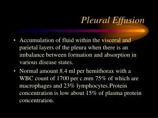
TRANSUDATIVE PLEURAL EFFUSION • Transudative pleural effusion are defined as effusion that are caused by systemic factors that alter the pleural equilibrium or starling forces • Components of starling forces : -Hydrostatic pressure - Permeability - Oncotic pressure
CAUSES OF TRANSUDATIVE PLEURAL EFFUSION Common causes Generalized salt and water retention • Congestive heart failure • Nephrotic syndrome Ascites • Cirrhosis • Peritoneal dialysis
Other Causes • Myxedema • Hypoalbuminemia • Urinothorax • CSF leakage • obstruction of brachiocephalic vein • Pulmonary veno-occlusive disease • Fontan procedure • Glomerulonephritis
Characteristics of Transudative Pleural fluid • Straw coloured • Non Viscous • Odorless • WBC < 1000 mononuclear predominant • Sugar > 60 mg/dl (except urinothorax ) • Ph 7.45-7.55
CONGESTIVE HEART FAILURE • Most common cause of Transudative pleural effusion • CHF accounts for 90 percent of transudative effusion • Usually small ,bilateral effusion • Unilateral effusion can occur ( 12 percent ) • More on right side by ratio of 2:1
PATHOPHYSIOLOGY CHF Increased Pressure in pulmonary capillaries Pleural effusion Increased amount of fluid enter the interstitial spaces of lung Increased interstitial pressure in subpleural interstitial spaces Fluid moves from the pulmonary interstitial spaces across the visceral pleura into pleural space
Signs and symptoms • H/O Increasing dyspnea on exertion • Increase in peripheral edema • Orthopnea or paroxysmal nocturnal dyspnea • : Dyspnea is frequently out of propotion to the size of effusion • Signs of both r/l side heart failure • Signs of pleural effusion
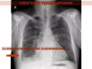
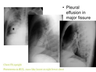
CHEST XRAY • Cardiomegaly and usually b/l pleural effusion
DIAGNOSIS If patient has • Cardiomegaly • b/l pleural effusion • Afebrile • No pleuritic chest pain • No need for thoracentesis • Treatment of CHF is started and observe the patient to determine whether the fluid is reabsorbed
INDICATIONS FOR THORACENTESIS • Effusions are not b/l and comparably sized • Pleural chest pain • Febrile • Effusions do not rapidly disappear after starting treatment of CHF
EFFECT OF THORACENTESIS IN CHF • Between the first and third thoracentesis , the mean protein level increased from 2.3 to 3.5 gm/dl • and the mean lactate dehyrogenase level increased from 176 to 262 IU/l
PRO BRAIN NATRIURATIC PEPTIDE • It based on ,when the ventricles are subjected to increased pressure or volume ,BNP is released • Biologically active BNP and larger amino terminal part N terminal probrain netriuratic peptide ( NT-ProBNP) are released in equimolaramounts in circulation PLEURAL FLUID (NT-ProBNP) • Levels below 100 pg/ml CHF unlikely • Levels above 1500 pg/ml Diagnostic of CHF
TREATMENT Aims of treatment • Decrease the venous hypertension • Control of heart failure • Treat heart failure with digitalis,diuretics • Therapeutic thoracentesis indicated in patient’s with heart failure and large pleural effusion refractory to treatment • In rare cases , pleurodesis with minocycline or tetracycline will be helpful.
NEPHROTIC SYNDROME • Nephrotic syndrome is a triad of proteinuria , hypoalbuminemia and edema • It is a non specific disorder in which the kidneys are damaged ,causing them to leak large amounts of protein (proteinuria at least 3.5 gm/day /1.7mtsquare body surface area ) from the blood into the urine. • Pleural effusion due to nephritocsyndrome are usually b/l and are frequently infrapulmonary in location • Pleural effusion occur in 1/5th of cases • Pleural effusion often associated with peripheral edema.
Pathophysiology Pleural effusion in nephrotic syndrome is due to combination of : • Decrease plasma oncotic pressure (because of protein loss) • Increase hydrostatic pressure (because of salt and water overload • Thromboembolic episodes of pulmonary vessels due to increase renal vein thrombosis or due to protein S deficiency may be another cause of pleural effusion
Investigations Diagnostic thoracentesis Should be perform to ascertain that the pleural fluid is transudate Lung scan or ct angiography should be done to rule out pulmonary embolism
Treatment Aims of Rx • Decreasing the protein loss in urine to increase the plasma protein • Decrease the increased extra cellular volume Rx • Diuretics • Low sodium diet • ACE inhibitors • Serial therapeutic thoracentesis should not be performed because they only further deplete the protein stores
HYPOALBUMINEMIA • Albumin ,the body’s predominant serum binding protein ,comprises 75-80 percent of normal plasma colloid oncotic pressure and 50 percent of protein content • Hypoalbuminemia caused by various conditions including nephrotic syndrome ,hepatic cirrhosis ,heart failure & malnutrition
According to starling equation hypoalbuminemia would decrease the oncotic pressure of the blood and increase the rate of pleural fluid formation • But study done in patients with different serum albumin levels , no significant difference of plasma oncotic pressure between patients with or without pleural effusion • Thus ,hypoalbuminemia is an uncommon cause of pleural effusion • The recognition of pleural effusion in patients of low serum albumin levels should prompt careful clinical evalution to identify the other potential causes for effusion .
CIRRHOSIS • Incidence of pleural effusion in cirrhosis of liver about 6 percent • Pleural effusion usually occur only when ascitic fluid is present • Pleural effusion in patients with cirrhosis and ascitis usually right sided (67 percent) left sided (16 percent )and b/l in (16 percent) • Right side is more involved because the right hemidiaphragm is more likely to have embryological developmental defects
Pathophysiology • Pleural effusion occur due to movement of the ascitic fluid from the peritoneal cavity into the pleural cavity • In patients with tense ascitis and increased intra abdominal pressure, the diaphragm may be stretched ,causing microscopic defects, the increased hydrostatic pressure in the ascitic fluid results in one way transfer of fluid from the peritoneal to the pleural cavity
Diagnosis • Both paracentesis and thoracentesis should be performed to ascertain that the ascitis and pleural fluid are compatible with the diagnosis and do not have high polymorphonuclear cell counts • Pleural fluid protein level is usually higher than the ascitic fluid protein level but is still below 3gm/dl &the pleural fluid LDH level is low • Occasionally blood tinged or frankly bloody pleural fluid is not much significant because it may occurs due to the patients poor coagulation status • Amylase level should be determine & cytological examination performed on both fluid specimens to rule out pancreatic ascitis or malignant disease .
In patients with cirrhosis, ascites, and pleural effusion there is possibility of spontaneous pleural infection, which is analogous to the spontaneous bacterial peritonitis • Spontaneous bacterial peritonitis diagnosed if they have cirrhosis along with a positive bacterial culture of the pleural fluid and a neutrophil count greater than 250 cells/mm3 or a negative culture and a neutrophil count greater than 500 cells/mm3. • Patients with spontaneous bacterial pleuritis have worse liver disease • The treatment of choice is an antibiotic to which the cultured bacteria are susceptible. • Tube thoracostomy is usually not indicated
Treatment • The management of pleural effusions associated with cirrhosis and ascites should be directed toward treatment of the ascites because the hydrothorax is an extension of the peritoneal fluid. • The patient should be put on a low-salt diet, and diuretics should be given. • The best diuretic therapy appears to be the combination of furosemide and spironolactone. • The initial starting dose is 40 mg of furosemide and 100 mg of spironolactone. • The doses can be increased up to 160 mg of furosemide and 400 mg of spironolactone daily. • Serial therapeutic thoracenteses are not indicated because the pleural fluid rapidly reaccumulates.
Other treatment options • Liver transplantation patients refractory to salt restriction and diuretics and remain symptomatic from the presence of the large pleural effusion. • Transjugular intrahepatic portal systemic shunt (tips). • Shunt are frequently ineffective because of the pressure differences between the peritoneal cavity, the pleural space, and the systemic veins. Because the pleural pressure is less than the central venous pressure, fluid will preferentially move to the pleural space rather than to the central veins. • Video-thoracoscopy with closure of the diaphragmatic defects and pleurodesis
Peritoneal Dialysis • Peritoneal dialysis is a well established means of renal replacement therapy. • Hydrothorax mostly on right side is a well known complication of peritoneal dialysis • It is observed in approximately 2 percent of all peritoneal dialysis patients
Pleural effusions can result from CAPD by the movement of the dialysate from the peritoneal cavity into the pleural cavity through a mechanism similar to that of cirrhosis and ascites. • The effusion developed within 30 days of initiating the dialysis in 50% of the patients, but 18% had been on dialysis for more than a year before the effusion developed. • The pleural effusion occurs on the right side approximately 90% of the time, but it can be bilateral or left sided.
Diagnosis : • Pleural fluid have glucose level intermediate between that of the dialysate and the serum. • Protein level below 3 g/dl • low LDH level (<100 IU/L) which is higher than that in the ascitic fluid.
Treatment : • Dialysis must be stopped • Dialysis should be stopped for 2 to 6 weeks • 50 percent of patients have resolution of hydrothorax after temporary cessation of PD for 2 – 6 weeks.
Other treatment options : • Pleurodesis : It should be done if there is recurrence after temporary cessation of PD. • VATS Diaphrgm can be examined for areas of leaking and can be clipped.
MYXEDEMA • Mxedema rarely presented by pleural effusion • Myxematous pleural effusion is more frequent in women • The cause of myxematous pleural effusion is increased capillary permeability with leakage of plasma proteins • Isolateral pleural effusion secondary to hypothyriodism can be either an exudate or a transudate • The diagnosis of myxedema should always be considered in older patients with pleural effusion when there are no pathognomic findings Treatment : effusions are resistant to treatment with digitalis and diuretics but disappear after treatment with thyroid harmone therapy
URINOTHORAX • Pleural fluid that accumulate due to retroperitoneal leakage secondary to urinary obstruction , trauma ,retroperitoneal inflammation,malignancy ,failed nephrostomy or kidney biopsy is called urinothorax • This is rare cause of pleural effusion • Pleural effusion develop within hours of the precipitating event and disappears rapidly once the obstruction is relived
Diagnosis : • Pleural fluid looks and smells like urine • Protein level < 1 gm/dl • LDH level high • pH below 7.2 but can be normal • Glucose can be normal or reduced Conformation of the diagnosis can be obtained with simultaneous measurements of pleural fluid and serum creatinine levels • Pleural fluid creatinine has been greater than the serum creatinine in all cases of urinothorax • Ratio higher than 1 and in most cases greater than 10
Brachiocephalic Vein obstruction • Obstruction of the brachiocephalic vein can lead to a pleural effusion. • Causes : • intrathoracic goiter • thrombosis of the vein due to a central venous catheter • hemodialysis catheter • pleural fluid is borderline transudate.
CSF leakage • Rare cause of pleural effusion. • Causes : • Ventriculopleural shunting, • Trauma • Diagnosis : • fluid is clear and colorless • protein level is very low. • Measurement of the pleural fluid b2 transferrin. • Diagnosis can be made by radionuclide cisternography.
Pulmonary Veno-Occlusive Disease • Rare disease • Characterized pathologically by evidence of repeated pulmonary venous thrombosis. • The characteristic histologic feature of pulmonary veno-occlusive disease is obstruction of pulmonary venules and veins by intimal fibrosis; intravascular fibrous septa are nearly always present. • The etiology of this disease is unknown.
Mechanism : • Increased interstitial fluid that results from obstruction of the pulmonary veins Sign and symptoms : • Slowly progressive dyspnea and orthopnaea • Signs of pulmonary hypertension • Pleural effusion in a patient with pulmonary hypertension is suggestive of the diagnosis of pulmonary veno-occlusive disease Diagnosis : • Pulmonary veno-occlusive disease can be definitively diagnosed by surgical lung biopsy. Treatment : • lung transplantation
Fontan Procedure • In Fontan procedure, the right ventricle is bypassed by an anastomosis between the superior vena cava, the right atrium, or the inferior vena cava and the pulmonary artery. • The procedure is typically performed for tricuspid atresia or univentricular heart. • Pleural effusion is a significant problem after the Fontan procedure.
The pathogenesis of the formation of the large amounts of pleural fluid postoperatively in these patients is not definitely known. • It is probably related to the increased systemic venous pressure.
Characteristic of pleural fluid : • Pleural fluid is transudate • Pleural fluid reveals primarily lymphocytes. Rx : • Diuretics • Fluid restriction • Ace inhibitors • Low fat diet • Oxygen Other treatment options : • Pleuroperitoneal shunt • Pleurodesis • Ligation of thoracic duct