Download

1 / 48
730 likes | 2.04k Views
Approach to Pleural Effusion. Dr Abdalla Elfateh Ibrahim Consultant & Assisstant Professor of Pulmonary Medicine King Saud University. Pleural Effusion. Pleural effusions are a common medical problem with more than 50 recognized causes including Local pleura disease

E N D
Approach to Pleural Effusion Dr Abdalla Elfateh Ibrahim Consultant & Assisstant Professor of Pulmonary Medicine King Saud University
Pleural Effusion Pleural effusions are a common medical problem with more than 50 recognized causes including Local pleura disease Underlying lung Systemic conditions Organ dysfunction Drugs It occur as a result of increased fluid formation and/or reduced fluid resorption.
Mechanism The pathophysiology of fluid accumulation varies according to underlying aetiologies. Increase permeability Increase pulmonary capillary pressure Decrease negative pleural pressure Decrease oncotic pressure Obstructed lymphatics
Types of pleural effusions Transudatespleural fluid proteins < 30 OR Exudatespleural fluid proteins >30
Causes of pleural effusion Transudates Very Common causes Heart failure Liver cirrhosis
Transudates Less Common causes Hypoalbuminaemia Peritoneal dialysis Hypothyroidism Nephrotic syndrome Mitral Stenosis
Causes of pleural exudates Common causes Malignancy Parapneumonic effusions Tuberculosis
Exudates Less Common causes Pulmonary embolism Rheumatoid arthritis and other autoimmune pleuritis Benign Asbestos effusion Pancreatitis Post-myocardial infarction Post CABG
Exudates Rare causes Yellow nail syndrome (and other lymphatic disorders ) Drugs Fungal infections
Clinical assessment and history Thorough history (Infection, malignancy , risk of PE , heart failure etc.) And physical examination.
History Drug history is important. Uncommon cause of exudative effusion (mesotruxate, Amiodarone Phenytoin, Nitrofurantoin and Beta- blockers ) >100 cases reported globally An occupational history Asbestos exposure and potential secondary exposure via parents or spouses should be documented.
Symptoms Asymptomatic Breathlessness Chest pain Cough Fever
Approximately 75% of patients with pulmonary embolism and pleural effusion have a history of pleuritic pain. Dyspnoea is often out of proportion to the size of the effusion Asymptomatic if it occupies less than a third of the hemithorax
Signs Decrease expansion Dull percusion node Decrease vocal resonance Decrease air entry Signs of associated disease (for example :chronic liver disease-CCF-nephrotic syndrome -SLE-RA-Ca lung)
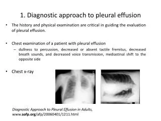
DIAGNOSIS CXR Pleural aspiration Pleural biopsy Medical thoracoscopy CT scan VAT Bronchoscopy
Pleural aspiration The initial step in assessing a pleural effusion is to ascertain whether the effusion is a transudate or exudate Diagnostic tap Aspiration should not be performed for bilateral effusions in a clinical setting strongly suggestive of a transudate, unless there are atypical features or they fail to respond to therapy
Pleural aspiration A diagnostic tap, with a fine bore (21G) needle and a 50mL syringe Bedside ultrasound guidance is recommended for all diagnostic aspirations Biochemistry : protein, LDH, PH, and glucose Microbiology: Gram stain, AFB and culture Pathology :cytology
Pleural aspiration Aspirated fluid should immediately be drawn into a blood gas syringe for PH Biochemical (2-5 ml) Microbiology 5ml 50ml for cytological examination
Pleural effusion Document in the patient file : Aseptic techique (under local Anathesia) The amount of effusion aspirated Appearance and odour should be noted. (colour usually Straw colour (normal) Smell , unpleasant aroma of anaerobic infection may guide antibiotic The appearance may be serous blood tinged or frankly bloody -
Appearance Milky fluid Empyaema Chylothorax PesudoChylothorax
Centrifuging turbid or milky pleural fluid will distinguish between empyema and lipid effusions. If the supernatant is clear then the turbid fluid was due to empyema If it is still turbid : -Chylothorax OR - Pseudochylothorax
Appearance Grossly bloody pleural fluid is usually due to Malignancy Pulmonary embolus with infarction Trauma Benign asbestos pleural effusions Post-cardiac injury syndrome
How to differentiate between haemothorax & hagic effusion • Pleural fluid haematocrit is greater than 50% of the patient's peripheral blood haematocrit is diagnostic of a haemothorax
Fluid Suspected disease Putrid odour Anaerobic empyema Food particles Oesophageal rupture Bile stained Cholothorax (biliary fistula) Milky Chylothorax/Pseudochylothorax ‘Anchovy sauce’ like fluid Ruptured amoebic abscess
Differentiating between exudate and transudate effusions Protein of > 30g/l an exudate Protein of <30 g/l a transudate. When protein is close to 30g/l (25-30)
Light's criteria Exudates if one or more of the following: Pleural fluid protein divided by serum protein is greater than 0.5 Pleural fluid LDH divided by serum LDH is greater than 0.6 Pleural fluid LDH > 2/3 the upper limits of laboratory normal value for serum LDH.
How accurate is Light’s criteria ? In CCF diuretic therapy increases the concentration of protein, LDH and lipids in pleural fluid In this context Light's criteria is recognized to misclassify a significant proportion of effusions as exudates . Clinical judgment should be used Measurement of NT-pro-BNP can be useful.
Other tests Glucose < 3.3 mmol/l ? Infection PH <7.2 empyaema Amylase pancreatic ca ,rupture oesophagus Rheumatoid factor RA ANA for SLE Complement level (reduced in SLE,RA,Ca)
Pleural fluid differential cell counts Cell proportions are helpful in narrowing the differential diagnosis but none are disease specific When any effusion becomes long standing it tends to be populated by lymphocytes (and neutrophils fade away) Pleural malignancy, cardiac failure and tuberculosis are common specific causes of a lymphocytic effusion
PH Pleural fluid pH should be measured in non-purulent effusions providing that appropriate collection technique can be observed and a blood gas analyser is available. Inclusion of air or local anesthetic in samples may significantly alter the pH results and should be avoided. In a parapneumonic effusion, a pH <7.2 indicates the need for tube drainage
PH In clinical practice, the most important use for pleural fluid pH is aiding the decision to treat pleural infection with tube drainage.
Pleural effusion cells (cont) Neutrophil (are associated with acute processes) Parapneumonic effusions: Pulmonary embolism Acute TB Benign asbestos related disease Eosinophils Pleural eosinophilia when eosinophyls aregreater than 10% of cells ( eosinophilic effusion) The most common cause eosinophilia is air or blood in the pleural space Is a fairly non-specific
Causes of lymphocytic p. effusions lymphocytes account for > 50% nucleated cells) Malignancy (including metastatic adenocarcinoma and mesothelioma) Lymphoma Tuberculosis
Causes of lymphocytic pleural effusions Cardiac failure Post CABG Rheumatoid effusion Chylothorax Uraemic pleuritis Sarcoidosis Yellow Nail Syndrome
Glucose In the absence of pleural pathology, glucose diffuses freely across the pleural membrane and pleural fluid glucose concentration is equivalent to blood A low pleural fluid glucose level (< 3.4 mmol/l) may be found in Complicated parapneumonic effusions Empyema Rheumatoid pleuritis Tuberculosis Malignancy Oesophageal rupture .
Glucose The most common causes of a very low pleural fluid glucose level (< 1.6 mmol/l) are Rheumatoid arthritis Empyema Although glucose is usually low in pleural infection and correlates to pleural fluid pH values, it is a significantly less accurate indicator for chest tube drainage when compared to pH
Cytology The diagnostic yield for malignancy depends on The skill and interest of the cytologist Tumour type. The diagnostic rate is higher for adenocarcinoma Than for Mesothelioma, Squamous cell carcinoma lymphoma and sarcoma.
Tumour markers Pleural fluid and serum tumour markers do not have a role in the investigation of pleural effusions.
Management Treatment of the cause Drainage (stop drain for 1-2 hours after 1st 1500 ml) may presipitate pul oedema Pleurodesis with - Talc - Tetracycline -Bleomycin Surgery