Download
1 / 45
450 likes | 456 Views
Biology 212 Anatomy & Physiology I. Digestive System. Functions:. Organs. Two functional groups: 1. Alimentary Canal or Gastrointestinal tract Organs which ingest, propel, digest, absorb, & eliminate Oral cavity, pharynx, esophagus, stomach,

E N D
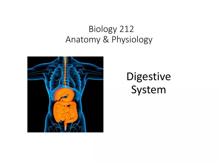
Biology 212Anatomy & Physiology I Digestive System
Organs Two functional groups: 1. Alimentary Canalor Gastrointestinal tract Organs which ingest, propel, digest, absorb, & eliminate Oral cavity, pharynx, esophagus, stomach, small intestine, large intestine, anus 2. Accessory Digestive Organs Assist with digestion Teeth, tongue, salivary glands, liver, gall bladder, pancreas
Oral Cavity: Includes: teeth tongue Opens posteriorly into oropharynx which leads to laryngopharynx Which opens inferiorly into esophagus
Paired Salivary Glands: Parotid Submandibular Sublingual
Saliva: 97% - 99% water Solutes: Ions (electrolytes) Antibodies Lysozyme (antibacterial) Amylase (enzyme – digests carbohydrates) Mucous Lubricates Cleans mouth and teeth Immune surveillance Begins carbohydrate digestion
Alimentary Canal: From esophagus to anus: 4 concentric layers or tunics Serosa or Adventitia Muscularis Externa Submucosa Mucosa Epithelium Lamina propria Muscularis mucosa
Esophagus: Propels food from pharynx to stomach Posterior to trachea & heart
Esophagus: Propels food from pharynx to stomach Posterior to trachea & heart Mucosa: Nonkeratinized stratified squamous epithelium Many mucous-secreting cells Thin lamina propria Thin muscularis mucosa Submucosa: Thick layer of dense irregular CT Muscularis Externa: Upper third = skeletal muscle Middle third = mixed Lower third = smooth muscle Adventitia: Thin layer of connective tisssue
Esophagus: Passes from thorax to abdomen through diaphragm, enters stomach (left of midline).
Stomach: Storage: Highly distensible Delivers chyme slowly to duodenum Secretion of mucous, hydrochloric acid Secretion of enzyme pepsin - Digestion of proteins Secretion of gastric lipase – Digestion of lipids Secretion of intrinsic factor – Absorption of B12
Stomach: Fundus Cardiac Region Pyloris Body
Stomach: Lesser Curvature Greater Curvature
Stomach: Mucosa thrown into folds called rugae
Stomach: Pyloric sphincter releases chyme slowly into duodenum
Small Intestine: Total length: 6 to 7 meters Diameter: 2.5 to 3 centimeters Duodenum Jejunum Ileum
Small Intestine: DUODENUM: ~ 25 cm long Receives secretions of liver (bile) pancreas (enzymes) Secretes mucous digestive enzymes bicarbonate – neutralize HCl JEJUNUM: ~ 2.5 m long Specialized for absorption ILEUM: ~ 3.5 m long Specialized for absorption
Small Intestine: Serosa or Adventitia Muscularis Externa Submucosa Mucosa Epithelium Lamina propria Muscularis mucosa
Small Intestine: Mucosa specialized three ways to increase surface area for absorption 3. 1. All layers of mucosa thrown into folds called plicae circulares
Small Intestine: Mucosa specialized three ways to increase surface area for absorption 2. Epithelium and lamina propria form finger-like villi 3.
Small Intestine: Mucosa specialized three ways to increase surface area for absorption 3. Plasma membranes of epithelial cells form finger-like microvilli 3.
Small Intestine: Mucosa specialized three ways to increase surface area for absorption 3. Nutrients, ions, etc. are absorbed through these plasma membranes and pass through the cells to the deeper lamina propria, where they are absorbed into capillaries & lymphatic vessels.
Large Intestine (colon): From ileocecal junction to anus ~ 2 meters long ~ 5 to 8 cm diameter Most digestion has already occurred (small intestine) Colon primarily absorbs water and electrolytes forms feces for elimination
Transverse colon Ascending colon Descending colon Sigmoid colon Cecum Rectum
Left colic (splenic) flexure Right colic (hepatic) flexure Sigmoid flexure
Abdominal Accessory Organs: Liver, Gall bladder, Pancreas, & associated ducts
Liver Most superior organ in abdomen Immediately inferior to diaphragm Partially protected by ribs Develops from embryonic intestine Mass ~ 1.5 kg Four lobes Blood supply: Hepatic artery from celiac trunk Hepatic portal vein from stomach intestine pancreas spleen
Liver Digestive functions: Regulate metabolism Store excess carbohydrate protein lipid Secrete bile: Produced from cholesterol Stored in gall bladder via cystic duct Transported to duodenum through common bile duct Emulsifies fats
Liver: Hepatic artery Hepatic portal vein Common bile duct Enter / exit together on inferior surface
Pancreas: Inferior & posterior to stomach Fits into concavity of duodenum Both endocrine (Insulin, glucagon) exocrine (digestive enzymes) Enzymes transported to duodenum by pancreatic duct (shares opening with common bile duct)
Pancreas: Produces many different digestive enzymes: Amylases: starches & monosaccharides glycogen & disaccharides Proteases: proteins amino acids Lipases: diglycerides & fatty acids triglycerides & glycerol Nucleases: nucleic acids nucleotides
The abdominal cavity is lined by a double-layered serous membrane called the peritoneum. Visceral layer – surrounds and is firmly attached to abdominal organs Parietal layer – attached to inner surface of body wall Between these = Peritoneal cavity
Mesentary – fold of peritoneum connecting visceral & parietal layers
More terminology: Intraperitoneal organs Retroperitoneal organs connected to body wall posterior to peritoneal by mesentaries cavity
Intraperitoneal organs have a serosa which includes the visceral layer of the peritoneum. Retroperitoneal organs are not covered by the visceral layer of the peritoneum, so the connective tissue on their external surface is an adventitia. Mesentery In reality, many retroperitoneal organs (no mesentery) have a serosa (visceral peritoneum) covering part of their wall and an adventitia (no visceral peritoneum) covering part of their wall.
IntraperitonealRetroperitoneal Stomach Duodenum Jejunum Ilium Cecum & Appendix Ascending colon Transverse colon Descending colon Sigmoid colon Rectum Liver Gall bladder Pancreas
Intestinal Motility: Moves liquid distally by peristalsis Mixes chyme from stomach with - Intestinal enzymes - Pancreatic enzymes - Bile from liver Increases contact of liquid with intestinal mucosa for absorption
Digestion: Breakdown of food into molecules small enough to be absorbed from the lumen of the intestine into its blood vessels. Four types of molecules = more than 95% of total: Water Sugars (carbohydrates) Amino acids Fatty acids Also: Nucleic acids Ions (electrolytes, minerals) Vitamins ? Others ?
Mechanical digestion In mouth (teeth) Chemical digestion Begins in mouth Continues through intestine Absorption of nutrients Primarily in electrolytes small intestine vitamins Absorption of water Both small intestine & large intestine Salivary glands produce amylases Stomach secretes proteases Small intestine secretes proteases Liver secretes bile Pancreas secretes amylases, proteases, lipases, nucleases
All absorbed molecules must pass through epithelial cells of intestinal mucosa. Tight junctions prevent anything from passing between cells.
Alimentary Canal: Since contents include many foreign substances, including microorganisms, your immune system considers it “outside” so Lined by many lymphatic and immune tissues in lamina propria and submucosa