Download
1 / 39
390 likes | 427 Views
Learn about the classifications and functions of joints, including fibrous, cartilaginous, and synovial joints. Explore the structural and functional aspects, range of motion, and key components of different joint types.

E N D
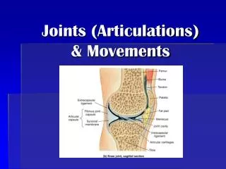
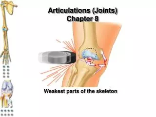
Joints (Articulations) - know • Articulation – site where two or more bones meet • Functions of joints • Give the skeleton mobility • Hold the skeleton together • Joints are a weak part of the skeleton and are often injured
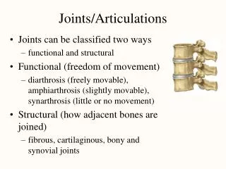
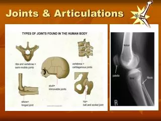
Classification of Joints: Structural - know • Structural classification focuses on the material binding bones together and whether or not a joint cavity is present • The three structural classifications are: • Fibrous (cranial bones) • Cartilaginous (ribs – sternum) • Synovial (knee)
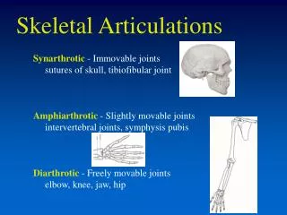
Classification of Joints: Functional - know • Functional classification is based on the amount of movement allowed by the joint • The three functional classes of joints are: • Synarthroses – immovable (cranial bones) • Amphiarthroses – slightly movable (vertebrae) • Diarthroses – freely movable (knee) We will discuss joints based on structural classification:
1. Fibrous Joints (structural classification) - know • The bones are joined by fibrous tissues • There is no joint cavity • Most are immovable There are three types – sutures, syndesmoses, and gomphoses
a. Fibrous Joints: Sutures - know • Occur between the bones of the skull • *Comprised of interlocking junctions completely filled with connective tissue fibers* • Bind bones tightly together, but allow for growth during youth • Skull bones fuse and are called synostoses • Begins as early as age 10-15, generally complete by age 30-50
Fibrous Structural Joints: Sutures - example Figure 8.1a
b. Fibrous Joints: Syndesmoses - know • *Bones are connected by a fibrous tissue ligament* • Movement varies from immovable to slightly variable • Examples include the connection between the tibia and fibula, and the radius and ulna
Fibrous Joints: Syndesmoses - example Figure 8.1b
c. Fibrous Joints: Gomphoses - know • The peg-in-socket fibrous joint between a tooth and its alveolar socket • The fibrous connection is the periodontal ligament
2. Cartilaginous Joints (structural classification) - know • Articulating bones are united by cartilage • *Lack a joint cavity* • Two types – synchondroses and symphyses
a. Cartilaginous Joints: Synchondroses - understand • A bar or plate of hyaline cartilage unites the bones • All synchondroses are synarthrotic (immovable) • Examples include: • Epiphyseal plates of children • Joint between the costal cartilage of the 1st rib and the sternum
Cartilaginous Joints: Synchondroses - example Figure 8.2a, b
b. Cartilaginous Joints: Symphyses - understand • Hyaline cartilage covers the articulating surface of the bone and is fused to an intervening pad of fibrocartilage • Amphiarthrotic (slightly movable) joints designed for strength and flexibility • Examples include intervertebral joints and the pubic symphysis of the pelvis (expansion in female for childbirth)
3. Synovial Joints (structural classification) - know • Most common • *Those joints in which the articulating bones are separated by a fluid-containing joint cavity* • All are diarthroses (freely movable) • Examples – all limb joints, and most joints of the body (regardless of size!)
Synovial Joints: General Structure - understand All synovial joints have: • Articular cartilage • Joint (synovial) cavity • Articular capsule • Synovial fluid • Reinforcing ligaments
Synovial Joints: General Structure - example Cadaver dissection
Synovial Joints: Friction-Reducing Structures - know These friction-reducing structures are found in synovial joints and are common where ligaments, muscles, skin, tendons, or bones rub together: • Bursae – flattened, fibrous sacs lined with synovial membranes and containing synovial fluid • Tendon sheath – elongated bursa that wraps completely around a tendon
Synovial Joints: Friction-Reducing Structures – understand complexity
Synovial Joints: Stability - understand Stability is determined by: • Articular surfaces – shape determines what movements are possible • Ligaments – unite bones and prevent excessive or undesirable motion • Muscle tone • Tendons of muscles cross the joint and help stabilize it • The tendons are kept tight by muscle tone
Synovial Joints: Movement - know The two muscle attachments across a joint are: • Origin – attachment to the immovable bone • Insertion – attachment to the movable bone • Described as movement along transverse, frontal, or sagittal planes
Synovial Joints: Range of Motion - know • Nonaxial – gliding movements only • Uniaxial – movement in one plane • Biaxial – movement in two planes • Multiaxial – movement in or around all three planes
1. Gliding Movements - understand • One flat bone surface glides or slips over another similar surface • Examples – intercarpal and intertarsal joints, and between the flat articular processes of the vertebrae
Angular Movement – know – (important slide!) • Flexion — bending movement that decreases the angle of the joint • Extension — reverse of flexion; joint angle is increased • Dorsiflexion and plantar flexion — up and down movement of the foot • Abduction — movement away from the midline • Adduction — movement toward the midline • Circumduction — movement describes a cone in space
Rotation – know (be able to identify) • The turning of a bone around its own long axis • Examples • Between atlas and axis • Hip and shoulder joints
Special Movements – know (be able to identify) • Supination and pronation • Inversion and eversion • Protraction and retraction • Elevation and depression • Opposition
Special Movements – know (be able to identify) Figure 8.6c
Special Movements – know (be able to identify) Figure 8.6d
How joints are classified Examples • Link to table of joints • Medical school site: Joints • Arthritis Foundation Website
Types of Joints: examples Various kinds of joints. Fibrous: A, syndesmosis (tibiofibular); B, suture (skull). Cartilaginous: C, symphysis (vertebral bodies); D, synchondrosis (first rib and sternum). Synovial: E, condyloid (wrist); F, gliding (radioulnar); G, hinge or ginglymus (elbow); H, ball and socket (hip); I, saddle (carpometacarpal of thumb); J, pivot (atlantoaxial).