Download
1 / 42
590 likes | 1.54k Views
The Biomechanics of Human Skeletal Articulations. Explain the functions of articular, fibro cartilage and articular connective tissue. Define joint stability and e xplain factors influencing its.

E N D
The Biomechanics of Human Skeletal Articulations Explain the functions of articular, fibro cartilage and articular connective tissue. Define joint stability and explain factors influencing its. Define joint flexibility and Explain the advantages and disadvantages of different techniques for increasing or maintaining joint flexibility. Describe the biomechanical contributions to common joint injuries and pathologies.
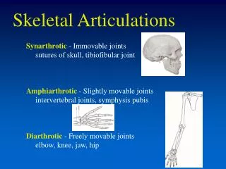
Classification of Joints based on Functions Immovable (Synarthroses) Absorb shock but permit little or no movement Slightly Moveable (Amphiarthroses) Freely Movable (Diarthrosesor Synovial) The articulating bone surfaces are covered with articular cartilage, an articular capsule and synovial membrane. Synovial membrane lining the interior articular capsule secretes a lubricant known as synovial fluid. Synchondroses – Sternocostal joint, epiphyseal plates Symphyses – Vertebral joints and the pubic symphysis • Sutures – Skulls • Syndesmoses - Tibiofibular joint
SUTURES • The only example in human body, the Skull. • Irregular grooved articulating bone sheets mate closely and are tightly connected by fibers.
Dense fibrous tissue binds the bone together SYNDESMOSES
AMPHIARTHROSES - SYMPHYSIS Thin plates of hyaline cartilage separate a disc of fibrocartilage from the bones.
SYNCHONDROSES Articulating bones are held together by a thin layer of hyaline cartilage
DIARTHROSES OR SYNOVIAL Articularcapsule consist of double layered membrane Synovial membrane (stratum) and Fibrous membrane. Synovial fluid -aclear, slightly yellow liquid that provides lubrication inside the articular capsule. Associated bursae - small capsules filled with synovial fluid that cushion the structures they separate
DIARTHROSES – CATEGORIZED BASED ON NUMBER OF AXES OF ROTATION PRESENT (a)Athrodial;(b)Sellar;(c)Ginglymus;(d)Trochoid;(e)Spherodial (f) Condyloid
Articular or Hyaline Cartilage Articular cartilage is a white connective tissue It covers the ends of bones.
FUNCTIONS OF ARTICULAR CARTILAGE What are the functions ofarticular cartilage? • It provides a protective lubricants that minimizes frictions and mechanical wear. • It spreads loads over a wide area, thereby reducing contact stress. • It has no blood supply and nerves. • Anisotropic, it deforms instantaneously to a low or moderate load. If rapidly loaded, it will become stiffer and deforms over a longer period.
ARTICULAR CARTILAGE • It is 1 to 5 mm thick, depending on the stress and the incongruity of the joint surfaces. • The force distributions depend on the cartilage thickness. Ankle and elbow, thin; hip and knee, thick. • It has a very low coefficient of frictions, range from 0.01 to 0.04, ice at 0º is about 0.1. Almost frictionless, allows gliding.
Transverse ligament Lateral meniscus Medial meniscus Anterior cruciate ligament Posterior cruciate ligament Superior view Functions of Articular Fibrocartilage What? It is a fibrocartilaginous disc or partial discs known as menisci that intervene between articulating bones.
ARTICULAR FIBROCARTILAGE Examples are the intervertebral disc and the knee joint. It is where both tensile strength and the ability to withstand high pressure are necessary.
What are the possible functions ofarticular fibrocartilage? • Distributing loads over joint surfaces • Protecting the joint periphery • Lubricating the joint • Absorbing shock at the joint • Improving the fit of articulations • Limiting slip between articulating bones
What are articular connective tissues? • Tendons - connect muscles to bones • Ligaments - connect bones to other bones Do not have ability to contract like muscle tissue but slightly extensible, elastic. Like bone it responds to mechanical stressby hypertrophying and atrophying. Therefore, regular exercises, increase size and strength of both tendon and ligaments.
What is joint stability? Ability of a joint to resist abnormal displacement (dislocation) of the articulating bones.
What factors effectjoint stability? • Orientation or joint position • Stability is maximal when joints are in the close-packed position(maximumcontact surface between the articulating bone). • Loosed-packed position- any joint orientation other than the closed packed position.
What factors effectjoint stability? Shape of the articulating surface • Acetabulum has relatively deep socket for the head of femur and large amount of contact area. • The small glenoid fossa has lesser contact area and diameter than humeral head, therefore contribute to relatively instable joint.
What factors effectjoint stability? A strong array of ligaments and muscle tendons crossing the joint Strong ligaments and muscle contributes significantly to joint stability by holding the articulating bone ends together (E.g. Strengthening the quadriceps and hamstring groups enhance the stability of the knee). The angle of attachment of most tendons to bones contributes to joint stability. The articulating ends of the bones at the joint are pulled closer together. Absence of muscle fatigue Since muscle strength effect joint stability, when muscle fatigues, joint stability also affected. Other Connective tissue Fascia and the bundles of muscle fiber within provide protections and support.
What isJoint Flexibility? • A description of the relative ranges of motion (ROM) allowed at a joint in different directions • ROM: the angle through which a joint moves from anatomical position to the extreme limit of segment motion in a particular direction. • Static flexibility – An indicator of the relative tightness or laxity of a joint. It refers to the ROM present when a body segment is passively moved (by an exercise partner or clinician). • Dynamic flexibility – It refers to the ROM achieve by virtue of muscle contraction.
Joint Flexibility Range of motion is measured directionally from anatomical position (zero).
What factors influencejoint flexibility? • Shape of the articulating bone surface and an intervening muscle tissue or fat terminate movement at extreme ROM. • Tightness in the muscle and collagenous tissue crossing a joint. When these tissues are not stretched, their extensibility usually diminishes (Collagenous tissue - extensibility increase with temperature elevation).
Joint Flexibility and Injury If the joint flexibility is extremely low or extremely high, the risk of injury is also increased. Too tight, possibility of tearing and rupturing are high. Too loose, it is prone to displacement-related injuries. People with less physical activity will have less flexibility. Stretching increase flexibility and avoid injury.
AGONIST AND ANTAGONIST Agonist The muscle that responsible for moving the body part to contracts or shortens. Antagonist The muscle that responsible for moving the body part back to its original position.
HOW TO INCREASE JOINT FLEXIBILITY? Stretching the ligaments and muscles that limit the ROM at joint will increase or maintain flexibility. Certain type of stretching being more effective than the other due to different neuromuscular responses. The proprioreceptors which transform mechanical distortion in the muscle or joint into nerve impulses that enter the spinal cord and stimulate the motor response.
NEURALMUSCULAR RESPONSE THAT INFLUENCING THE JOINT FLEXIBILITY Golgi Tendon Organs (GTO) What? It is a sensory receptors that influence extensibility. Where? It is located in the muscle-tendon junction and in the tendons at both ends of muscles. How? It is stimulated by tension either produce by muscle contraction or by passive muscle stretch.It inhibit tension in agonist muscle (promote relaxation) & initiate tension development in antagonists.
GTO MECHANISM Contractions of quadriceps muscle increases the tension in the patellar tendon which stimulates the Golgi tendon organ. The impulse travel to the spinal cord and some of them head upward to the higher centers of the nervous system for further interpretation and integration. The remaining messages excite either, interneurons that inhibit quadriceps function (extension of the knee), or interneurons that activate hamstring function (flexion of the knee).
NEURALMUSCULAR RESPONSE THAT INFLUENCING THE JOINT FLEXIBILITY Muscle Spindles What? It is a sensory receptors that are respond to the amount of muscle (static response) and rate of muscle lengthening (dynamic response). Where? It is interspered throughout and parallel to the fibers of muscles. How? Muscle spindles will provoke reflex contraction in stretched muscle & inhibit tension in antagonists..
KNEE JERK TEST A tap on the patellar tendon initiates the stretch reflex, resulting in the jerk caused by the immediate tension development in the quadriceps group.
KNEE JERK TEST MECHANISM Tapping the tendon stretches the quadriceps femoris muscle group. This activates stretch receptors within the muscle called muscle spindles. Stretching a spindle fiber initiates a volley of impulses in the sensory neuron (called an "I-a" neuron) attached to it. Impulse from muscle spindle travel to spinal cord. Some impulse are carried back to the same muscle causing it to contract. The leg straightens.
KNEE JERK TEST MECHANISM Some of the branches of the I-a axons synapse with inhibitory interneurons in the spinal cord. These, in turn, synapse with motor neurons leading back to the antagonistic muscle that inhibits tension development in antagonist muscle.
Golgi Tendon Organs and Muscle Spindles: How do they Compare? Muscle Spindles Interspersed among muscle fibers in parallel with the fibers Increase in muscle length 1) initiate rapid contraction of stretched muscle, 2) inhibit tension development in antagonist muscles Golgi Tendon Organs Within tendons near the muscle-tendon junction in series with muscle fibers Increase in muscle tension 1) inhibit tension development in stretched muscle, 2) initiate tension development in antagonist muscle Promote stretch in muscle being stretched Location Stimulus Response Overall Effect Inhibit stretch in muscle being stretched
BETWEEN GTO’S AND MUSCLE SPINDLE GTO promotes relaxation in muscle developing tension, whereby Muscle Spindles inhibit stretch in muscle being stretch. Therefore, maximizing GTO and minimizing the muscle spindle effect is the general goals in increasing joint flexibility
Techniques for Increasing Joint Flexibility What are active and passive stretching? • Active stretching isproduced by contraction of the antagonist muscles (Quadriceps contract so that Harmstring is stretched). It provides the advantage of exercising the muscle group used to develop force. • Passive stretchingisproduced by a force other than tension (Gravitational force, force applied by another body segment or by another person) in the antagonist muscles. Movement can be carried out father beyond the ROM (compare to active stretching)
Techniques for Increasing Joint Flexibility What areballisticandstatic stretching? • Ballistic stretching -a series of quick, bouncing-type stretches. The potential for injuries is heightened. • Static stretching- maintaining a slow, controlled, sustained stretch over time-usually about 30 seconds. This type of stretching is preferred because ballistic activate the muscle spindle respond that inhibits stretching. Why static stretching did not activate muscle spindle?
Techniques for Increasing Joint Flexibility What is PNF? Proprioceptive neuromuscular facilitationis a group of stretching procedures involving alternating contraction and relaxation of the muscles being stretched
COMMONT JOINT INJURIES AND PATHOLOGIES Sprains – abnormal displacement or twisting of the articulating bones results in stretching or tearing of ligaments, tendons, and connective tissues crossing joints.
DISLOCATION Dislocation - Displacement of the articulating bones at a joint. Result from fall or involving large magnitude of force. Symptoms include pain, swelling and loss of joint movement capability.
BURSITIS Bursitis – Overuse injury caused by excessive use of a joint that produces frictional irritation and inflammation of one or more bursae. Pain and some swelling are symptoms of bursitis.
ARTHRITIS Arthritis - an inflammation of a bone joint accompanied by pain and swelling. Rheumatoid Arthritis- Type of arthritis where body’s autoimmune system attack healthy tissue. the synovium becomes inflamed. This inflammation causes chemicals to be released that thicken the synovium and damage the cartilage and bone of the affected joint. This leads to inflammation of the synovium causing pain and swelling
Osteoarthritis What is osteoarthritis? • a common, degenerative disease of articular cartilage • symptoms include pain, swelling, ROM restriction, and stiffness • cause is unknown • both too little and too much mechanical stress seem to promote development
Cartilage that cushions the bones of the hip starts to erode, eventually allowing the bones to grind or rub together and causing hip pain and stiffness.The exact cause of osteoarthritis is unknown.