Download
1 / 43
430 likes | 456 Views
Welcome to Anatomy & Physiology II course with Dr. Greg Erianne. Learn about blood, plasma, hemostasis, and more. Access course materials, syllabus, and exam study guides online. Follow suggested study methods for success. Develop biomedical terminology mastery and logical thinking skills. Get ready for lectures, discussions, and lab sessions.

E N D
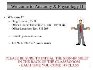
Welcome to Anatomy & Physiology II • Who am I? • Greg Erianne, Ph.D. • Office Hours; Tues/Fri 9:30 am – 10:30 am • Office Location: Rm. SH 205 • E-mail: gerianne@ccm.edu • Tel; 973-328-5377 (voice mail) PLEASE BE SURE TO INITIAL THE SIGN-IN SHEET IN THE BACK OF THE CLASSROOOM - EACH TIME YOU COME TO CLASS
Emergency Evacuation Procedures • Emergency evacuation may be required when there is an actual or potential danger to the occupants of any building as a result of fire or other emergency situation. When a fire alarm is sounded, all occupants must leave the building(s) via the nearest exit and proceed immediately to the designated staging area and remain 50 feet from any building. Fire Marshals will direct the evacuation. All walkways and roads must remain clear for emergency vehicles. Take all belongings with you. You will remain there until the all clear is sounded, or a Fire Marshal directs you to a remote staging area. Evacuation of physically disabled individuals will be assisted or coordinated by the faculty at the site. DO NOT USE ELEVATORS DURING THE EVACUATION PROCESS. The evacuation staging area for this classroom or laboratory is: • DH First Floor (lecture); • Primary: Rear exit to lot 1 50 ft past walkway • Secondary: Parking lot 1 • SH Second Floor (lab); • Primary: Lawn above HH stairs • Secondary: Parking lot 5
Overview of Lecture 1 • Course and Publisher Web sites • Course Description/Textbook/Lab Book • Course Objectives and Syllabus Review • Blueprint for success/Study strategy • Overview of blood • Blood volume and composition • Formed elements of blood • Blood plasma • Hemostasis • Blood groups and transfusions
Course Web Sites • Our Web sites for this class are located at: • http://www.gserianne.com/science/GerianneBio102/ (Main) • Announcements (VERY IMPORTANT TO LOOK AT FREQUENTLY!) • Syllabus and all lecture schedules • Lecture slides used in class (ppt and pdf formats) • Supplementary online materials for Lecture (and Lab – check with your instructor) • Lecture Exam Study Guides • Links to many other sites including Pearson’sWeb site • http://courses.ccm.edu (Blackboard Learn; Secondary) • You will need your student ID and password for the Blackboard (BB) site • This BB site will be used ONLY grades and grade-related things • http://masteringaandp.com (from Pearson Science) • You will need the course ID and have to register if you haven’t been to this site before • Lots of resources to use for A&P II – take advantage of it! • (Course ID: MAPERIANNE01390) • Printing slides and other materials (see email I sent)
Outline of Course/Requirements • Course Description • Lecture / discussion format • Lectures may not follow the order of Marieb’s Human Anatomy & Physiology, 10th edition – please check your syllabus! • Figures used for class • Laboratory • Marieb’s Laboratory Manual, 12th edition • Reading assignments for lab should be done BEFORE you come to lab
Blueprint for Success • Most importantly… • Skim your textbook BEFORE lecture and make notes • Take notes in your own words and become mentally involved during lecture; review/rewrite your notes after lecture • Ask questions if you don’t understand • Continually review previously learned material • Use all the study aids available to you • ***Before taking the exam, you should be able to take a BLANK study guide and answer all the questions WITHOUT YOUR NOTES!!!! • **See the Suggested Study Method on Web at the gserianne.com Web site – Please review this!!! • **Be sure to print slides/materials if you want them for class/lab – make a schedule for yourself – don’t get behind!
Major objectives of this course • In general, you will… • Master the objectives listed in the syllabus • Develop a further mastery of scientific/biomedical terminology • Further develop your ability to think logically and critically • Let’s review the syllabus and handouts…
Marieb’s Human Anatomy and Physiology Marieb w Hoehn Chapter 17 Blood Lecture 1
Overview of Blood – (Hem(o)-) Blood is what type of tissue? Connective tissue. Functions • transports vital substances (O2, waste) • maintains stability of interstitial fluid • distributes heat • hemostasis • prevents infection Blood Cells (formed elements) • form in red bone marrow • red blood cells • white blood cells • platelets (cell fragments) Plasma (liquid portion - matrix) • contains dissolved substances • mostly water and proteins • amount of blood varies with • body size • changes in fluid volume • changes in electrolyte concentration • amount of adipose tissue • about 7-8% of body weight • About 5.0 liters of blood in adult
Blood Composition Hematocrit (HCT) – Percentage of red cells in blood by volume. Also called Packed Cell Volume (PCV). Usually about 45% What would happen to the hematocrit if someone was dehyrated and lost plasma volume? Figure from: Saladin, Anatomy & Physiology, McGraw Hill, 2007
Formed Elements of the Blood 45% of blood Figure from: Hole’s Human A&P, 12th edition, 2010
Origin of Blood Cells All formed elements of blood arise from a common hematopoietic pluripotent stem cell (a hemocytoblast) in the red bone marrow Figure from: Hole’s Human A&P, 12th edition, 2010
Red Blood Cells • erythrocytes • biconcave (↑ surface area) • one-third hemoglobin(~ 280 million Hb molecules per RBC) • oxyhemoglobin • deoxyhemoglobin • can readily squeeze through capillaries • lack nuclei and mitochondria Figure from: Hole’s Human A&P, 12th edition, 2010
Hemoglobin General structure: - Four polypeptides chains - A porphyrin - An iron atom Heme Figure From: Martini, Anatomy & Physiology, Prentice Hall, 2001
Red Blood Cell Count • number of RBCs in a cubic millimeter (mm3) of blood. (1 mm3 = 1 microliter, µl) • 4,600,000 – 6,200,000 in males • 4,200,000 – 5,400,000 in adult females • 4,500,000 – 5,100,000 in children Average is about 5 x 10⁶ RBCs / µl • Number of RBCsreflects blood’s oxygen carrying capacity
Red Blood Cell Production • low blood oxygen causes kidneys and liver to releaseerythropoietin which stimulates RBC production(up to 30 million per second under maximum EPO stimulation!) • Erythropoiesis • vitamin B12, folic acid and iron necessary for RBC production Figure from: Hole’s Human A&P, 12th edition, 2010
Blood Viscosity and Osmolarity • Viscosity (thickness) • Resistance to flow of blood • Whole blood is about 5x as viscous as water • Changes in viscosity can put strain on the heart • Erythrocytosis (polycythemia) viscosity • Osmolarity • Due to NUMBER of ‘particles’ dissolved, not the type • Na+, proteins, erythrocytes • Osmolarity determines fluid flow between blood and tissues
Red Blood Cell Turnover The average life span of an RBC is about 120 days (4 months) Iron is carried in the blood by transferrin to red bone marrow, liver Figure From: Martini, Anatomy & Physiology, Prentice Hall, 2001 Porphyrin from worn out RBCs is converted into biliverdin and bilirubin
Types of Anemia Anemia – deficiency of RBCs or Hb in RBCs; reduces O2-carrying capacity of blood • aplastic anemia • bone marrow damaged • toxic chemicals • radiation • iron deficiency anemia • hemoglobin deficient • lack of iron • pernicious anemia • excess of immature RBCs • inability to absorb B12 • hemolytic anemia • RBCs destroyed • toxic chemicals • thalassemia • hemoglobin deficient • RBCs short-lived • defective gene ( or -chain) • sickle cell anemia • abnormal shape of RBCs • defective gene (-chain)
White Blood Cells • leukocytes • protect against disease • interleukins and colony-stimulating factors stimulate development in red bone marrow • granulocytes • neutrophils • eosinophils • basophils • agranulocytes • lymphocytes • monocytes ‘phils’ are filled with granules!
Neutrophils • light blue granules in acid-base stain • lobed nucleus • other names • segs • polymorphonuclear leukocyte (PMNs) • bands (young neutrophils) • first to arrive at infections • phagocytic • *55% - 65% of leukocytes (most numerous type of WBC) • elevated in bacterial infections Figure from: Hole’s Human A&P, 12th edition, 2010
Basophils Figure from: Hole’s Human A&P, 12th edition, 2010 • deep blue granules from basic stain • release histamine andheparin in allergic reactions (similar to mast cells) • less than 1% of leukocytes
Eosinophils Figure from: Hole’s Human A&P, 12th edition, 2010 • deep red granules in acid stain • bilobed nucleus • participate in allergic reactions • defend against parasitic worm infestations • 1% - 3% of leukocytes • elevated in worm infestations and allergic reactions, collagen diseases, diseases of spleen
Monocytes Figure from: Hole’s Human A&P, 12th edition, 2010 • largest blood cell • agranulocyte • kidney-shaped or oval nuclei • leave bloodstream to become macrophages • 3% - 9% of leukocytes • elevated in typhoid fever, malaria, tuberculosis, viral infections, inflammation
Lymphocytes • about the size of RBC • agranulocytic • large spherical nuclei • thin rims of cytoplasm • T cells • B cells • NK cells • important in immunity • produce antibodies • 25% - 33% of leukocytes • decreased T Cells in AIDS Figure from: Hole’s Human A&P, 12th edition, 2010
Diapedesis • Diapedesis - leukocytes squeeze through capillary walls to enter tissue space outside the blood vessel Figure from: Hole’s Human A&P, 12th edition, 2010
White Blood Cell Counts • number of WBCs per mm3 of blood • 5,000 – 10,000 per mm3 (or μl) of blood • leukopenia(-penia = deficiency of cell number) • low WBC count • typhoid fever, flu, measles, mumps, chicken pox, AIDS • leukocytosis(-cytosis = increase in cell number) • high WBC count • acute infections, vigorous exercise, great loss of body fluids • differential WBC count • lists percentages of types of leukocytes • may change in particular diseases
Origin of Blood Cells All formed elements of blood arise from a common hematopoietic pluripotent stem cell (a hemocytoblast) in the red bone marrow Figure from: Hole’s Human A&P, 12th edition, 2010
Blood Platelets • called thrombocytes when nucleated (in birds) • cell fragments of megakaryocytes • membrane bound • 150,000 – 500,000 per mm3 of blood (average ≈ 350,000 per µl) • help control blood loss from broken vessels
Blood Plasma • straw colored • liquid portion of blood • 55% of blood Figure from: Hole’s Human A&P, 12th edition, 2010
Plasma Proteins Albumins • most numerous plasma proteins (~55%) • ‘transport’ proteins • originate in liver • help maintain osmotic pressure of blood Alpha and Beta Globulins • originate in liver • transport lipids, metal ions, and fat-soluble vitamins Gamma Globulins • originate inlymphatic tissues (plasma cells) • constitute the antibodies of immunity Fibrinogen • originates in liver • plays key role in blood coagulation
Gases and Nutrients Gases • oxygen • carbon dioxide • nitrogen Nutrients • amino acids • simple sugars • nucleotides • lipids • lipoproteins
Non-protein Nitrogenous (NPN) Substances • molecules containing nitrogen that are not proteins • urea – product of protein catabolism; about 50% of NPN substances ( BUN – blood urea nitrogen; one indicator of kidney function) • uric acid – product of nucleic acid catabolism • amino acids – product of protein catabolism • creatine – stores phosphate groups (energy) • creatinine – product of creatine metabolism
Plasma Electrolytes • sodium • potassium • calcium • magnesium • chloride • bicarbonate • phosphate • sulfate
Hemostasis • cessation of bleeding Platelet Plug Formation • triggered by exposure of platelets to collagen • platelets adhere to rough surface to form a plug Blood Vessel Spasm • triggered by pain receptors, platelet/endothelial cell release of various substances • smooth muscle in vessel contracts (vascular spasm) Blood Coagulation • triggered by platelets, cellular damage and blood contact with foreign surfaces • blood clot forms 1. Vascular phase(Blood vessels) 2. Platelet phase(Platelets) 3. Coagulation phase(Plasma clotting factors Hemostasis
Platelet Plug Formation Substances released by platelets: - ADP (platelet activator) - thromboxane A2 and serotonin (vessel constriction) - clotting factors - Ca2+ (aids in coagulation) - PDGF Example of positive feedback Figure from: Hole’s Human A&P, 12th edition, 2010
Blood Coagulation Three cascades: • Instrinsic • Extrinsic • Common * Coagulation is an example of positive feedback ~ 15 sec. ~ 3-6 min. Figure from: Martini, Anatomy & Physiology, Prentice Hall, 2001
Blood Coagulation Extrinsic Clotting Mechanism (shorter, faster) • chemical outside of blood triggers blood coagulation • triggered by tissue factor (III) or thromboplastin (not found in blood, thus it’s extrinsic and produced by damaged tissue) Intrinsic Clotting Mechanism (longer, slower) • chemical inside blood triggers blood coagulation • activators are in direct contact with blood or contained within the blood triggered by Hageman factor (XII; found inside blood) • triggered when blood contacts a ‘foreign’ surface, e.g., collagen fibers, glass tube Both pathways are activated after blood vessel damage
Blood Clots • After forming, blood clot retracts (~60%) and pulls the edges of a broken vessel together • Platelet-derived growth factor stimulates smooth muscle cells and fibroblasts to repair damaged blood vessels • Thrombus – blood clot (in undamaged vessel) • Embolus – blood clot moving through blood Serum is the fluid expressed from a clot, i.e., the plasma minus clotting factors
Factors Preventing Coagulation • The smooth lining (endothelium) of blood vessels discourages the accumulation of platelets • Prostacyclin released by endothelial cells(aspirin) • Some cells secrete heparin (an anticoagulant) • As a clot forms, fibrin absorbs thrombin and prevents the reaction from spreading • Antithrombin (in plasma) interferes with the action of excess thrombin (activated by heparin) • Plasmin digests blood clots (generated from plasminogen via the action of a plasma enzyme, kallikrein, and tPA) *
Review To remember relative % of leukocytes from most to least numerous: “Never Let Monkeys Eat Bananas”