Download
1 / 32
320 likes | 476 Views
Joints (arthritis) – Rheumatoid arthritis. Inflammatory dz affecting synovial joints predominately Hyperplasia of synovial fibroblasts Severity is varied Peak age is 30-50 About 1% of the population is affected with a 2.5xs higher risk in women. May be genetic as it tends to run in families

E N D
Joints (arthritis) – Rheumatoid arthritis • Inflammatory dz affecting synovial joints predominately • Hyperplasia of synovial fibroblasts • Severity is varied • Peak age is 30-50 • About 1% of the population is affected with a 2.5xs higher risk in women. • May be genetic as it tends to run in families • More common in Native Americans
Insidious over several weeks usu. • Usu. Not symmetric at the beginning • Must have an inflammatory synovitis • If deformity is in a non-wt bearing jnt, you can assume it’s due to synovitis • 85% have serum RF • May start sero-(-) but become sero(+) w/ progression • ESR is typically helpful to follow the inflammatory activity
Prolonged morning stiffness is universal • Active phase: warm, swollen jnts Structural damage • Bone on bone crepitus • Try injected corticosteroids for anti-inflammation • C/s – neck stiffness w/ possible loss of motion • C1 transverse lig tenosynovitis and possible z-jnt synovitis • Pain doesn’t always accompany instability even in significant myelopathy
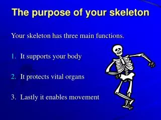
Immobilization due to pain is the kiss of death to joint mobilizations. The result is contractures and deformity • You need to keep pt’s ROM esp. in non-wt bearing jnts like shoulder and hand • Once cartilage is completely gone, bones may fuse if immobilized10% remit usu in first two yrs of dz • 90% of jnts that are affected are involved during the 1st yr • Severe dz= 10-15yr decr in life expectancy due to infection, pulmonary or renal dz, lymphoproliferative disorders, GI bleeds and cardiovascular
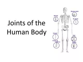
RA criteria below: • Morning stiffness • 3+jnts • Arthritis of hand • Symmetric arthritis • Rheumatoid nodules • RF • X-ray changes • Need 4 of the 7 • 1-4 must occur for at least 6 wks
Stage I Early – no destruction • Stage II Moderate – no jnt deformity, osteoporosis w/ or w/out some bone and cartilage destruction • Stage III Severe – cartilage and bone destruction with osteoporosis, jnt deformity • Stage IV Terminal – fibrous or bony ankylosis
Remission criteria • 5+ of the following for 2+ consecutive mos. • Morning stiffness</=15minutes • No fatigue • No jnt pain • No tenderness • No swelling • ESR
GOUT • Monosodium urate deposition - hyperuricemia • Tophi – accumulation of crystal in articular, osseous, ST, and cartilage • Recurrent attacks of inflammation • Uric acid calculi in GU; renal fxn impairment called gouty nephropathy • M/c 5th decade men African-Americans • Serum urate levels rise over time in men but don’t in women until after menopause due to estrogen • Gout in women is often due to thiazide diuretic use and renal failure • Blacks due to more HTN, not genetic
Crystals have decr solubility in low temps that’s why it likes toes and ears • Likes areas of minor trauma like 1st MTP • Hemiplegia – tophi won’t form on paralyzed side -> something to do w/ CT structure and turnover • Tophi is inflammatory cells around crystal w/ erosion of surrounding cartilage and bone. Fibrous capsule around tophi • Crystals are needle-shaped and formed radially
Three stages: asymptomatic hyperuricemia, acute intermittent gout, Chronic tophaceous gout. • Initially rubor, tubor, dolor and pain of jnt. Pain incr. Over hours. Pt. May not be able to walk. May get fever, chills, malaise. May last up to 2 weeks. Attacks become more frequent w/ time • ½ involve 1st MTP as monoarticular site and 90% of pts overall
CHRONIC TOPHACEOUS GOUT • About 10yrs after initial dx usu. • No pain free period but not as severe as acute • Factors for tophi development: early onset, long active phases, 4+attacks/yr, UE or polyarticular episodes • Tophi can also be in heart valves and sclera • Supcutaneous gouty tophy are usu in fingers “heberden’s nodes”
Early onset gout • <25yoa, 3-6% of gout pts, 80% have FHx • More severe and rapid course, 25% have nephrolithiasis • Transplant gout • 75-80% of heart transplants • Due to cycloporine tx to prevent rejection • Inhibits urate excretion
10% die of renal failure; 25% have renal stones • 25-50% have HTN ->due to reduced renal blood flow from urate • Hyperlipidemia/obesity – contraversial • Xray – ST swelling -> asymmetric in peripheral jnts erosions slightly removed from jnt (unique) (“overhanging edge”) • No osteopenia, and maintained jnt space until late
OA • Garrod in 1907 differentiated RA from OA10% of OA patients had reduced work hours and 13.7% retired early. • Arthritis is the main reason for decr. Activities in the elderly