Download
1 / 117
1.19k likes | 2.48k Views
Rheumatoid Diseases of the Hand. R. Dale Reynolds, M.D. UT Houston Plastic and Reconstructive Surgery. Rheumatoid Arthritis. A 58 yo man requests definitive relief of severe wrist pain. A radiograph is shown. Which of the following is most appropriate: Arthroplasty with silicone implant

E N D
Rheumatoid Diseases of the Hand R. Dale Reynolds, M.D. UT Houston Plastic and Reconstructive Surgery
Rheumatoid Arthritis • A 58 yo man requests definitive relief of severe wrist pain. A radiograph is shown. Which of the following is most appropriate: • Arthroplasty with silicone implant • Intercarpal arthrodesis • Proximal row carpectomy • Radial shortening • Total wrist arthrodesis
Rheumatoid Arthritis • 53 yr old woman with RA has Stage IV disease of the MCP. Which is the most characteristic posture of the patient’s MCP and PIP?
Rheumatoid Arthritis • 49 yr old woman with RA of wrists and hands has loss of active extension of the MCP of R middle and ring fingers for the past 6 months. PE shows full passive ROM of MCP; x-rays show no joint subluxation. When the digits are passively extended, she is able to maintain extension against resistance. Which is the most appropriate next step in management? • Observation • Arthroplasty of MCP of middle and ring • Repair of extensor tendon ruptures of middle and ring • Centralization of the extensor tendons of the middle and ring fingers at the MCP joints • Arthrodesis of MCP of middle and ring fingers
Rheumatoid Arthritis • The photograph shows a 60 year old man with advanced RA who has a boutonniere (type I) deformity of the thumb. Which of the following is the most likely cause of his findings? • Rupture of the EPL tendon • Rupture of the FPL tendon • Tenosynovial proliferation at the carpometacarpal joint of the thumb • Tenosynovial proliferation at the IP joint of the thumb • Tenosynovial proliferation at the MCP joint of the thumb
Rheumatoid Arthritis • 42 yr old woman with severe RA has advanced joint degeneration, pain, and decreased use of the right elbow, wrist and hand. On PE, the elbow is stiff and tender and the wrist and MCP joints are tender and subluxed. Radiographs confirm these findings. Which of the following staged sequences id the most appropriate? • Elbow arthroplasty, wrist arthrodesis, MCP joint arthroplasties • Elbow arthroplasty, MCP joint arthroplasties, wrist arthrodesis • MCP joint arthroplasties, elbow arthroplasty, wrist arthrodesis • MCP joint arthroplasties, wrist arthrodesis, elbow arthroplasty
Rheumatoid Arthritis • Changes in last 10 years • More aggressive treatment • Better drugs and medical management • Fewer isolated synovectomies • More severe deformities • Etanercept and Infliximab • Further radical change soon
Rheumatoid Arthritis • Diagnosis (4 of 7) • > 1 hr morning stiffness x 6 weeks (PA) • > 2 simultaneous PA regions x 6 weeks • Hand joint arthritis x 6 weeks • Symmetric arthritis x 6 weeks • RA nodules on extensor tendons (physician) • + Serum RF • Erosion or decalcification on x-rays (PA)
Rheumatoid Arthritis • May be initial presenting problem • Destructive synovitis • RA synovitis is associated with angiogenesis and a high lymphocyte content • Aggregates • Tendon sheath and synovial joints • Limited motion or rupture • Joint deformity
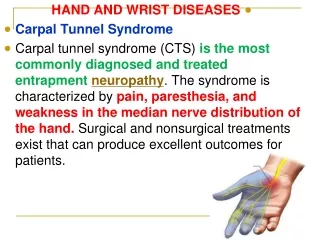
Rheumatoid Arthritis • Wrist, MCP, PIP • RA nodules • Carpal tunnel syndrome • Digital vasculitis • Ischemic neuropathy • Muscle wasting • Raynaud’s phenomenon
Rheumatoid Arthritis • Autoimmune process • 50-80% + RF (IgM) against IgG • 15% normal population + RF • Seronegativity (Rheumatoid variants) questionable surgical prognosis
Rheumatoid Arthritis • High ESR in active phase • Plain x-rays (PA swelling, erosions) • U/S more sensitive for PA swelling (80%) • Synovial fluid (differential, crystals) • Gout, calcium pyrophosphate disease • Synovium in all joints and portions of flexor and extensor tendons (beneath pulleys)
Rheumatoid Arthritis • Stages • Proliferative: Swelling, pain with motion, limited movement, nerve compression • Destructive: Synovial erosion causes irreversible changes (tendon rupture, capsular weakness and disruption, bone erosion, joint subluxation and deformity) • Reparative:Fibrosis replaces inflammation (adhesions, ankylosis, fixed deformity)
Rheumatoid Arthritis • History • Monocyclic (10%) – One attack • Polycyclic (45%) – Variable duration, severity and intervals • Progressive (45%) - Unremitting
Rheumatoid Arthritis • History • Ask which aspects give them the most trouble • Often very functional although deformed • Functional grading • No incapacity • Manages all but the heaviest tasks • Only lightest duties • Chair or bed bound
Rheumatoid Arthritis • History • Pain • Recent increase in pain may indicate a “flare-up” • Not usually present at rest (only exacerbations) • Demonstration of painful movement can help localize most active joints • Stiffness • How long does it take to loosen up in the morning (limber up time =LUT)
Rheumatoid Arthritis • History • Numbness and paraesthesias • Compression: Median nerve more common than in general population due to increase synovial volume • Generalized neuropathy: Common in the lower limb • Weakness • From pain, joint collapse, synovitis • Appearance • Can be main complaint
Rheumatoid Arthritis • History • Medications • Steroids – wound healing, stress dose • Synovectomy during proliferative phase should wait until 3-6 month medical trial
Rheumatoid Arthritis • Non-articular effects • Iritis or uveitis - 3-5% • Scleromalacia perforans - globe rupture due to nodule necrosis • Anemia – 25% (normocytic, normochromic) • Polyneuropathy – lower limbs • Cardiac – pericarditis, myocarditis, valvular • Arteritis - uncommon
Rheumatoid Arthritis • Non-articular effects • Lung changes • Rheumatoid lung: Honeycombed appearance on CXR due to multiple nodules • Caplan’s syndrome: Found with pneumoconiosis massive pulmonary fibrosis • Idiopathic pulmonary fibrosis
Rheumatoid Arthritis • Rheumatoid syndromes • Felty’s syndrome: RA + LAD + splenomegaly granulocytopenia / anemia • Splenectomy has no effect on arthritis • Sjogren’s syndrome (2 of 3): RA + keratoconjunctivitis sicca, xerostomia
Rheumatoid Arthritis • Examination • Neck, shoulder, elbow, radius, ulna, wrist extensors, thumb, MCP, PIP, DIP, nerve compression • Usually symmetrical joint involvement • Dominant hand often more advanced
Examination Deformities on general inspection Posterior subluxation of elbow Palmar subluxation of wrist Ulnar translocation of carpus Radial deviation of the wrist Ulnar drift of the fingers Palmar subluxation of MCP Swan neck or boutonniere deformity of fingers Z- deformity of the thumb Lateral dislocation of any of the IP joints Misalignment of digits suggestive of tendon rupture Rheumatoid Arthritis
Rheumatoid Arthritis • Z mechanism • Characteristic of many deformities of rheumatoid hand • Joint adopts an angulation in one direction, the joints on either side will tend to go in the opposite direction • Due to changes in mechanical advantage of tendons acting on a series of joints
Rheumatoid Arthritis • Gross Instability (Arthritis Mutilans, Opera- Glass Hands) • Results when bone ends are excessively eroded • Flail joints result • Grasp becomes impossible • Ankylosis of a joint may arrest its shortening and cause a disproportionately long digit • Arthrodesis of all affected IP joints early with bone grafting where appropriate
Rheumatoid Arthritis • Swellings • Rheumatoid nodules: Firm and rubbery (swollen olecranon bursa is fluctuant), most at ulnar border just distal to elbow but can be anywhere, poor prognosticator, painful, may ulcerate, should be excised • Benign pseudorheumatoid nodules: Histologic not clinically equivalent
Rheumatoid Arthritis • Swellings • Rheumatoid nodulosis: Seropositive adults, histologically rheumatoid, mild arthralgia, mild radiographic changes • Prominent ulnar head • Synovial swelling
Rheumatoid Arthritis • Skin • Thin, papery especially with steroids • Bruising, petechiae, fingertip hemorrhage, infarct • Psoriasis – elbow, fingernails, seronegativity • Palmar erythema – thenar and hypothenar • Intertrigo – accumulation of moisture most often b/w fingers and in the palm with MCP
Rheumatoid Arthritis • Muscle wasting • Excessive thenar wasting suggests possible median nerve compression • First dorsal interosseous suggested by deep concavity of dorsal aspect of the first web space
Rheumatoid Arthritis • Systemic regional assessment • Joint by joint, tendon by tendon, nerve by nerve • Pain • Synovial swelling • Tenderness • Range of motion (active, passive, associated pain) • Stability • Crepitus • Deformity (fixed, mobile)
Rheumatoid Arthritis • Neck (common) • Atlanto-axial subluxation • Superior migration of odontoid into foramen magnum • Subaxial subluxation of the vertebral bodies • Pain, instability, neurological disturbance • Full assessment before any general anesthetic (passive ROM, trigeminal nerve testing, lower limb reflexes)
Rheumatoid Arthritis • Neck • Neural deficit • I: Nil • II: Subjective weakness, hyperreflexia, dysaesthesia • IIIa: Objective long tract signs • IIIb: Quadriparesis • All should have AP and lateral in flexion and extension • Only 1 % need posterior fusion (II or III)
Rheumatoid Arthritis • Shoulder • Tested by touching interscapular region • Difficulty requires evaluation by orthopedic surgeon
Rheumatoid Arthritis • Elbow • Synovium – Active synovitis causes swelling • ROM – Normal 0-145o , synovitis causes pain at extremes, synovectomy help pain but not always ROM • Crepitus – Creaking or grinding with passive motion indicates marked synovitis or erosion • Stability – Dislocation uncommon, posterior, severe instability assessed by evaluation at 90o (Fig. 5.16)
Rheumatoid Arthritis • Radial head • Evaluate abducted at 90o supporting, pronating and supinating • Crepitus – Loss of articular cartilage in superior radioulnar joint, excision of the radial head is indicated and improves pain and ROM • ROM – Normal pronation 70o, supination 85o • Pain – Extreme supination often causes pain at ulnar head
Wrist Synovitis Extensor compartment: Ulnar>radial Hour glass constriction on exam from retinaculum Joints of wrist: Can be divided into three compartments each lined with synovium (radiocarpal, mid-carpal, radioulnar) Cartilage degradation, synovial expansion, ligamentous laxity Rheumatoid Arthritis
Rheumatoid Arthritis • Cartilage destruction • Chemical effects of intra articular lysosomes and free oxygen radicals • Synovial expansion • Essence of RA • Bony erosions bony spicules / tendon rupture • Attempted grip increases pressure and pain
Rheumatoid Arthritis • Ligamentous laxity • Stretching from distended synovium • Carpal supination and ulnar translocation and associated with scapholunate dissociation • Muscle tendon units crossing deform carpal collapse palmar facing carpal row / carpal height / ROM / strength • Degenerative arthritis due to bony contact • Carpal supination and palmar subluxation of the distal radius and carpus dorsal subluxation of the distal ulna in the region of the distal radioulnar joint
Rheumatoid Arthritis • Ligamentous laxity • All may be affected but ulnar carpal complex (UCC) is commonly first • Synovitis causes: • Scalloping laterally • Eventual rupture of the triangular fibrocartilaginous complex (TFCC) distally and can damage extensors 4 and 5 with ulnar head erosion • Threatens effectiveness of ECU medially
Rheumatoid Arthritis • Ligamentous laxity • Radioscaphocapitate (RSC) or sling ligament is second • Joined by incompetence of the radioscapholunate (RSL) rotatory subluxation of the scaphoid / loss of radial carpal height • Joined by radiolunatriquetral (RLT) and wrist subluxes into anterior position
Rheumatoid Arthritis • Ligamentous laxity • Dorsal radiocarpal(DRC) ligament fails ulnar translocation of carpus • If whole carpus moves then type I translocation • Mid-carpal joint is relatively spared usually • Triquetrohamitatocapitate (THC) can deteriorate volar intercalated segment instability (VISI)
Rheumatoid Arthritis • Ligamentous laxity UCC prominent ulnar head supinated carpus ineffective ECU RSC +RSL loss of radial height + RLT + DRC ulnar translocation = radial carpal rotation anterior wrist subluxation (Shapiro angle = 112o in normal, higher in RA) [radial cortex index metacarpal to line from tip of radial styloid to the ulnar corner of the distal radius]
Rheumatoid Arthritis • Ligamentous laxity • Prominent ulnar head extensor tendon rupture • Radial carpal rotation changing alignment of the metacarpus and encouraging ulnar drift (Z) • Anterior wrist subluxation reduced efficiency of the extrinsic tendons weakens grasp / encourages swan neck deformity
Rheumatoid Arthritis • PE • Synovium • Three finger test for fluctuance over ulnar / dorsal • ROM • Normal: Flexion 75o, extension 70o, ulnar deviation 35o, radial deviation 20o • Pain • Synovitis extremes of range • Articular cartilage loss limited arcs
Rheumatoid Arthritis • Distal Radioulnar Joint (Ulnar Head) • Inherently unstable • Sigmoid notch never contacts more than 60% of the ulnar head • In some positions it is <10% • Stability largely from TFCC but ECU, pronator quadratus, the interosseous membrane, the dorsal carpal ligaments and FCU all contribute