Download
1 / 44
460 likes | 520 Views
Explore different types of cardiomyopathies, their causes, clinical presentations, diagnostic investigations, and treatment modalities including medical and non-pharmacological approaches. Enhance your knowledge for better patient care.

E N D
Cardiomyopathies Prof. Lotfy Hamed Abo dahab. Professor Of Internal Medicine & Cardiology. Sohag University.
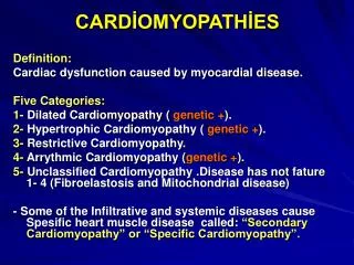
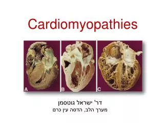
Cardiomyopathy • Def: • Cardiomyopathy is a general term indicating disease of • the cardiac muscle. • Classification: • Cardiomyopathies are classified according to the • predominant clinical presentation into: • 1- Dilated cardiomyopathy- ventricular dilation. • 2- Hypertrophic cardiomyopathy- myocardial hypertrophy. • 3- Restrictive cardiomyopathy- impaired vent. filling. • 4- Arhythmogenic right ventricular cardiomyopathy (ARVC)-right ventricular involvement is prominent • with a high frequency of ventricular arrhythmias.
Dilated Cardiomyopathy • Def: • DCM is characterized by dilation and impaired systolic function of LV and/ or RV, in the absence of abnormal loading conditions (e.g. HTN, valve dis.) and coronary artery diseases. • Causes of DCM • 1- Genetic: • - Autosomal dominant DCM. • - X-linked DCM. • 2- Inflammatory: • - Post infective • - Autoimmune • - Connective tissue dis.(SLE,scleroderma.)
3- Metabolic: e.g. Glycogen storage dis. 4-Nutritional: e.g. Thiamine def. 5- Endocrinal: - Acromegaly - Thyrotoxicosis - Myxedema - DM 6-Infiltrative: Hereditary haemochromatosis. 7-Neuromuscular: - Muscle dystrophy - Frederic's ataxia.
8-Toxic: - Alcohol - Cocaine - Adriamycin. 9-Haematological: - Sickle cell anemia - TTP. 10- Puerperal cardiomyopathy. 11- Ischemic cardiomyopathy.
Clinical presentation: • 1- Congestive heart failure: • - Symptoms and signs of left and/or right heart failure. • 2-Syncope or sudden cardiac death: • - Due to ventricular arrhythmias, or conduction defect. • 3-Pulmonary or systemic embolism. • Clinical evaluation should include careful family history and evaluation of relatives.
Investigations: 1-Chest X-ray:Shows cardiomegaly 2-ECG:- Sinus tachycardia - Non specific ST,T changes - Conduction abnormalities and arrhythmias. 3-Echocardiography: - Dilation of LV and/ or RV with poor global contraction. 4-Coronary angiography: - Should be performed to exclude coronary artery dis. in all pts. at high risk.
Treatment:- • Goals of treatment: • 1- To relieve symptoms. • 2- To retard disease progression. • 3- To prevent complications. • Lines of treatment: • 1- Conventional mangement of heart failure: • Diuretics - ACE-inhibitors - ARBs • B-blockers • 2-Mangement of arrhythmias: • - Anti-arrhythmic drugs - Permenant pacing • - Implantable cardioverter-defibrillators.
3-Anticoagulation treatment: is indicated in: - Severe ventricular dilation and dysfunction, - Documented atrial fibrillation, or - History of embolization. 4-Cardiac transplantation: The principal option for refractory HF, and advanced dis. 5- Alternatives to cardiac transplantation: -Ventricular assist devices (Extracorporeal devices, implantble pulsatile devices, total artificial heart). -Intra-aortic ballon counterpulsation. -Cardiomyoplasty.
Hypertrophic cardiomyopathy • Def.: Hypertrophic cardiomyopathy (HCM) is defined as myocardial hypertrophy in the absence of an identifiable cause. • Prevalence: The prevalence of HCM is about 1 in 500. • Patho-physiology: - There is disarray of cell to cell arrangement, disorganization of cellular architecture and fibrosis. - The most common sites of ventricular involvement are the septum, apex and mid ventricle.
Clinical presentation: • Signs and symptoms:1) Heart failure: • - Dyspnea on exertion • - PND and fatigue (diastolic HF).2)Myocardial ischemia3)Syncope and presyncope : • - As a low COP symptom • - Associated with increased risk of SD.4)Sudden death: • - It is more common among children and young adults, • - It occurs mainly during physical activity. Both arrhythmias and ischemia can be the underlying mechanisms.
Investigations:1) ECG: • - LVH • - RVH • - RA , and LA enlargement.2)Echocardiography: • - RVH • - LVH and may be obstruction • - Diastolic dysfunction.
Treatment of HCM:-Aims of treatment:1-Mangement of heart failure (diastolic and systolic HF).2-Prevention of SD.I-Medical therapy:1-B-blockers: Are effective in reliving angina, dyspnea and syncope.2-Calcium channel blockersII-Non-pharmacological therapy:1- Septal myomectomy2- ICD or amiodarone to prevent SCD.
Def: Restrictive cardiomyopathies are group of diseases characterized by:- Restricted filling function - Increase atrial pressure - Dilatation of the atria (Thrombus formation commonly occur) - Congestive heart failure When the dis. Advances, LV contraction is decreased. Restrictive cardiomyopathy
● Classification:1- Primary restrictive cardiomyopathy:- Idiopathic restrictive cardiomyopathy - Endomyocardial fibrosis - Esinophilic cardiomyopathy2- Secondary restrictive cardiomyopathy - Amyloidosis - Hemochromatosis - Sarcoidosis - Storage diseases - Radiation therapy (myocardial fibrosis)
●Clinical presentation:I- At the beginning pts. Presented with manifestations of diastolic dysfunction as : 1- Dyspnea on exertion and decreased exercise tolerance. 2- Dyspnea at rest, PND, and orthopnea. 3- Congestive heart failure: - Oedema - Rt. Upper abdomen pain - Abdominal bloating - Increased JVP,….II- Then manifestations of systolic heart failure present as: - Low COP …………..
Investigations:1- Chest x-ray:- Pulmonary venous congestion. - Atrial enlargement. - Cardiomegaly.2- ECG:- Low voltage. - Nonspecific ST segment and T wave abnormalities.3- Echocardiography:Will reveal diminished diastolic function of the heart .4- Cardiac catheterization:Help distinction from constrictive pericarditis
5- Endomyocardial biopsy:May be needed to reveal the cause of myocardial restriction. ●Treatment: -Treatment of heart failure. - Treatment of embolic manifestations. - Treatment of 1ry amyloidosis: Melphalan + prednisolone + Colchicine - Cardiac transplantation.
Arrhythmogenic right ventricular cardiomyopathy ● Def: It is a primary myocardial disease that mainly affects the RV and later on both ventricles are affected by progressive fibrofatty replacement of the Rt. ventricular myocardium. ●Histopathology: There is an evidence of patchy programmed cell death (apoptosis) of cardiac myocytes, and myocytes are replaced by fat tissue.
Clinical presentation:ARVC usually present itself among youngs as: - Syncope - Sudden death (due to ventricular arrhythmias) - Or more rarely as right sided heart failure. - Rare form of ARVC is associated with dermatological abnormalities (Naxos disease) caused by mutation in a gene encoding a myocyte structural protein (Plako globin)
Investigations:1- Chest x-ray: • - No abnormalities except in advanced disease. • 2- ECG: • - T wave inversion in Rt. Ventricular precordial leads • (V1 – V3). • - Small wave occurring at the end of QRS complex • (epsilon wave). • - RBBB. • 3- Holter monitoring: • - Demonstrates frequent extrasystolies (Rt. Ventricular origin) and runs of ventricular tachycardia.
4- Echocardiography: - May show areas of decreased RV contractility - Later on dilatation of the RV.5- MRI:- Helps to identify fatty infiltration of the RV.6- RV angiography: 7- RV biopsy: 8- Genetic testing:
Treatment: 1-Antiarrhythmic drugs: - β-blockers. - Amiodarone. 2- ICD. 3- Cardiac transplantation.