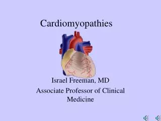
CARDİOMYOPATHİES
CARDİOMYOPATHİES. Definition: Cardiac dysfunction caused by myocardial disease. Five Categories: 1- Dilated Cardiomyopathy ( genetic + ). 2- Hypertrophic Cardiomyopathy ( genetic + ). 3- Restrictive Cardiomyopathy. 4- Arrythmic Cardiomyopathy ( genetic + ).

CARDİOMYOPATHİES
E N D
Presentation Transcript
CARDİOMYOPATHİES Definition: Cardiac dysfunction caused by myocardial disease. Five Categories: 1-Dilated Cardiomyopathy ( genetic+). 2- Hypertrophic Cardiomyopathy ( genetic +). 3- Restrictive Cardiomyopathy. 4- Arrythmic Cardiomyopathy (genetic+). 5- Unclassified Cardiomyopathy .Disease has not fature 1- 4 (Fibroelastosis and Mitochondrial disease) - Some of the Infiltrative and systemic diseases cause Spesific heart muscle disease called: “Secondary Cardiomyopathy” or “Specific Cardiomyopathy”.
Secondary Myocardial Diseases Definition: Myocardial disease of known origin. (Secondary Cardiomyopaty) 7 Subgroups: 1- Ischemic Cardiomyopathy 2- Hypertensive Cardiomyopathy 3- Valvular Cardiomyopathy 4- Alcoholic Cardiomyopathy 5- Metabolic Cardiomyopathy 6- Muscular Distrophy Cardiomyopathy 7- Peripartum Cardiomyopathy
HYPERTROPHİC CARDİOMYOPATHY (HCM) Definition:Massive ventricular hypertrophy with no absolute cause. • Not all of these patients show IHSS (Idiopatic Hypertrophic Subaortic Stenosis) or HOCMP (Hypertrophic Obstructive Cardiomyopathy) features. • There is no pressure gradient at left ventricle outflow(LVOT) tract in 1/3 of patient. (Rest or provocated gradient). • HCMP prevalance: 1/500. HCMP is the most prevalant genetic cardiovascular disease. • Nearly %60 inherited. Relation with mutation of beta-myosin heavy chain and also other sarcomeric proteins has been shown. (Troponin- T, -I, -C, Myosin Related Protein- C etc.). • HCM ıs the most frequent cause of sudden cardiac death among young people including athletes.
Structure of Human Sarcomere:Contraction is carried out by the interaction of actin and myosin. The process begins with the ligation of calcium to troponin complex (I, C, T) and alpha-tropomyosin. Then, actin binds to myosin and activates ATPase of the spheric myosin, so that contractile function is carried out. Cardiac myosin binded protein C binds to myosin and starts contraction.
HYPERTROPHIC CARDIOMYOPATHY: Cellular changes of myocytes. Cell disarrangement ise seen on microscope. Left: Organized and parallel cell arrangement in normal myocardium. Right: Disarrangement affects impuls conduction and promotes ventricular arrythmias.
Common HCM types:(A)- LV free wall, diffusely hypertrophied İVS. (B)- Asymmetric septal hypertrophy (C)- MassiveConcentric LV hpertrophy ( involve walls, papillary muscles). (A)- (B)- -(C)
HCMP: Patophysiology • Most patients have septum hypertrophy with non-dilated LV and/or RV cavity. Hypertrophy of the septum may be diffuse or only the upper, mid or apical hypertrophy of the septum may take place. • Hypertrophy is extednded to the LV free wall in most of the patients. • Diastolic filling is impaired because of the incomplete relaxation and compliance of the LV. • Hypertrophic LV empties most of its content in the first half of systole. (Hyperdynamic systolic function”). • Mitral anterior leaflet is displaced towards septum during mid systole and this causes obstruction the the LV outflow tract during this period. • At the obstructive phase of the disease mitral regurgitation is always present. There ise resting pressure gradient at LV outflow tract in %35 of patients. Gradient can be precipitted at the other %25 (by augmenting myocardial contraction, and reducing ventricular volume). • Fibrosis and occlusive disease in in small coronary arteries. The major coronary arteries, are wide and patent unless occlusive atherosclerosis occurs.
HCM: Symtomps:Angina, Dyspne, Presyncope, Syncope. Physical Findings: • 1- Before LV beat, LA beat is palpated (S4): Is present even in the absence of any gradient or murmur. Impaired LV relaxation. • 2- LVOT Sistolik ejectıon Murmur: Crescendo-Decrescendo, starts with S1 and ends with S2. Best heard between apex and left sternal border. Cervical radiation is weak. Augmented by manouvers and drugs which decrease preload. (Valsalva, standing, amyl nitrite). Attenuates with increasing afterload (squating, handgrip fenilefrin). • 3- MR murmur: Heard at late systole, radiates to axilla, and related with LV outflow obstruction. Mitral diastolic rumble and Paradoxic splitting of S2 may be heard. • 4- Hyperdynamic carotis pulse.
HCM: Clinical Presentation and Mechanisms • Chest pain: Ischemia, LVOT ob. Reduced coronary perfusion pressure. • Exertional dyspnea: Diastolic dysfunction. • Reduced functional capacity: LVOTob, systolic dysfunction, AFwith uncontrolled rapid ventricular rate. • Palpitation: SVT, AF, frequent VPB, non-sustained VT. • Syncope/Presyncope: Supraventricular arrythmia, LVOTob, vasovagal, high VT rate . İnadequate increase cardiac output during the effort. • Cardiac arrest: VT, SVT, AF, VF, bradyarrythmia.
HCM: Sudden death: Supraventricular arrythmia is present in %20-50 of the patients, but sudden death is caused by ventricular arrythmia. Major Risk Factors of Sudden Death: 1- Patient who survive after cardiac arrest (sustained ventricular tachycardia). 2- Arrythmia and abnormal blood pressure response to excersize. 3- Non-sustained ventricular tachycardia (Holter). 4- Family history of sudden cardiac death and syncope in more than two first degree relatives before 40 years. 5- LV Wall thickness >30 mm.
HCM: Other manifestations.Atrial Fibrillation • Seen in %15 of patients with HCM. • Absence of atrial systole. Rapid ventricular rate causes pulmonary edema or hypotension. • Rapid ventricular rate causes detoriation of functional capacity. • By conversion to sinus rythm or decreasing heart rate functional capacity improves. Endocarditis: May occur on aortic or mitral valves. Unexpected heart failure and IE symptoms or signs should be suggest İE in HCM patiernts.
HCM: ECG and CHEST FİLM Chest film: May be normal-large left heart chambers. . No aortic calcification. ECG: Is anormal in %97 of symptomatic HCMP, and in %90 of asymptomatic HCMP patients. • AF is detected in %15. • Non-sustained VT is frequent. . • Q waves in DII, DIII, aVF and D1, aVL, V5, V6 (and less frequently in V1-3). This sign shows hypertrophy, and causes pseudoinfarct patern. • Intraventricular conduction delay. High voltage findings of LVH. T waves of LVH. Huge negative T waves are frequently seen in apical HCMP high precordial QRS voltage. • Short PR and pre-exitation may be seen, but is infrequent.
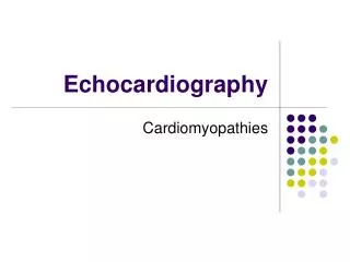
HCMP: Echocardiographic Hallmarks • Asymetric (dispropotionate) septal thickening: Septum to posterior wall ratio > 1.5 • LV myocardial segment >1.5 cm in thicknesss. • Poor Septal contractıon. Hypercontractile free posterior wall. • Systolic anterior motıon of the mitral valve (SAM) when outflow tract gradient >30 mmHg . • Mid-systolic closure of aortic valve. • Small LV cavity. • Mitral regurgitation is frequent. • LVOT gradient at rest present in about %35 of patients
HCM: Characteristic ECG paterns. • Left axis deviation • LBBB • Pathologic Q wave on anterolateral leads. • T wave inversion (commonly in İnferolateral leads) • ST segment changes. • Criteria for left atrial enlargement. • V3-5 or V4-6 huge T wave inversion (“Distal- apikal HCM”
OBSTRUCTIVE PHASE: Beta bockers: (Especially latent obstruction). Verapamil. Disopiramid.Amiodaron:(SVT,VA + ). Digoxin: (contraindicated). Diüretic: (contraindicated). ACEI (contraindicated). Surgery: Septal myectomy. Septal ablation. ICD is indicated in severe VA. LAST PHASE: Beta blockers: (small doses) Verapamil:Contraindicate. Disopiramid:Contraindicate. Digoxin:Beneficial. Diuretic: Beneficial. ACEI: Beneficial in patients with LV dilatation and HF. Surgery: Transplantation. HCMP: Management. Pharmacologic and surgical intervention.
Hypertrophic obstructive cardiomyopathy (HOCM): As Mitral valve changes:When the LVOTis narrowed, blood rushes through the passageway toward the aortic valve(like tight garden hose nozzle), dragging the leaflets of the mitral valve with it. Mitral valve normally functıons keep blood floıwing in direction from the left atrium (upper heart chamber) to the LV. However increased force of blood caused by HCM pulls the valve open and may cause blood leak backward (called regurgitatıon )into the LA. NORMAL LVOTobs.
Anterıor replacement of the papillary muscle in HOCM : MR, SAM
HOCM: Septal myectomy: Before the operatıon There is severe hypertrophy of the basal septum, whith systolic anterior motıon of the mitral valve (-A-). This results in severe LVOT obs. as well as MR. During the surgery (-B ), yhe portıon of the basal septum that project into the outflow tract is removed by scalpel, resulting in abolitıon of the LVOTobs. (-C-). There is no longer SAM, and theMR abolished.
HOCM:Septal ablation (with absolute ethanol). İndication: LVOT gradient at rest > 30-50 mmHg. With provocation 75-100 mmHg.