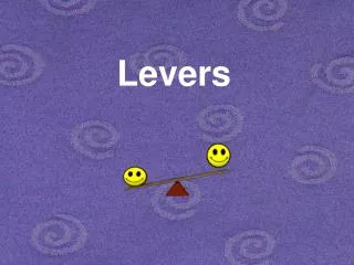
Muscles as Levers
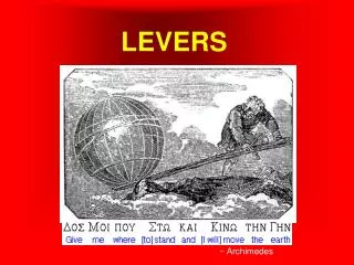
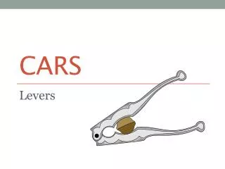
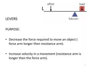
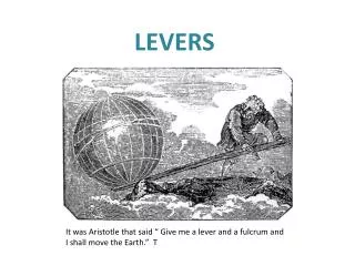
Muscles as Levers. Bones and muscles interact as levers : A simple machine used to transmit force A lever has 4 parts: Rigid bar/ rod Fulcrum or pivot where the bar turns Object moved against resistance Force that supplies energy for the bar to move Types: 1 st class lever

Muscles as Levers
E N D
Presentation Transcript
Muscles as Levers • Bones and muscles interact as levers: A simple machine used to transmit force • A lever has 4 parts: • Rigid bar/ rod • Fulcrum or pivot where the bar turns • Object moved against resistance • Force that supplies energy for the bar to move • Types: • 1st class lever • 2nd class lever • 3rd class lever
First Class Lever • Fulcrum is located between the resistance and the force (resistance – fulcrum - force) • Ie. Scissors, seesaw, and hemostats • Ie. Tilting the head
Second Class Lever • Resistance is located between the fulcrum and the force (fulcrum- resistance- force) • Ie. Wheelbarrow • Ie. Standing of the your toes
Third Class Lever • Force is located between the force and the resistance (fulcrum- force- resistance) • Ie. Tweezers
Types of Levers • You will be rotating about the room to different stations. • At each station you will be given a different task. • After completing the task you must determine the type of lever each of the following actions requires.
Origin & Insertion • Origin – attachment to an immoveable bone • Insertion – attachment to a movable bone • ILIOCOSTALIS= attaches to the ilium & ribs (costal = ribs)
Direction of Muscle Fibers • Relative to the Midline • RECTUS = parallel to the midline • RectusAbdominus • TRANSVERSE = perpendicular to midline • TransverseAbdominus • OBLIQUE = diagonal to midline • External Oblique
Location • Structure near which muscle is found • FRONTALIS = near FRONTAL bone • OCCIPITALIS = near OCCIPITAL bone
Size • Relative Size of Muscle • MAXIMUS = largest • GluteusMaximus • MEDIUS = middle • Gluteus Medius • MINIMUS = smallest • Gluteus Minimus • LONGUS = longest • FibularisLongus • BREVIS = short • FibularisBrevis • TERTIUS = shortest • FibularisTertius
Number of Origins • Number of tendons of origin • BICEPS = Two • Biceps Brachii • Biceps Femoris • TRICEPS = Three • Triceps Brachii • QUADRICEPS = Four • Quadriceps Femoris
Shape • Relative Shape of the Muscle • DELTOID = triangular shape Δ • TRAPEZIUS = trapezoid shape SERRATUS = saw-toothed ♒ • RHOMBOIDEUS = rhomboid shape • TERES = round ○
Anatomy of the Muscular System • Origin • Muscle attachment that remains fixed • Insertion • Muscle attachment that moves • Action • What joint movement a muscle produces • i.e. flexion, extension, abduction, etc.
Movement of Muscle • Muscles can only pull to create movement • Muscles rarely work alone, & are usually arranged in groups surrounding a joint: • Agonist (Prime Mover)- muscle that contracts to create action (major responsibility for certain movement) • Synergist- muscle that helps the agonist (aids the agonist/ prevents rotation) • Antagonist-muscle that opposes the agonist(undoes the desired action)
Types of Muscle Contraction • For each of the sporting examples, you must do the following: • Research the types of muscles that are used in the example • Determine the agonist and antagonist muscles • Determine the contraction type
Muscles of Facial Expression • Epicranius: covers the upper cranium; consists of 2 parts: • Frontalis- covers frontal bone • Occipitalis- covers occipital bone United by epicranialaponeurosis(tendinous muscle) that contracts to raise the eyebrows thus wrinkling the forehead
Muscles of Facial Expression • OrbicularisOculi- muscle encircling the eye; closes the eye & compresses the lacrimal gland • OrbicularisOris- encircles the mouth; helps close and pucker the lips
Muscles of Facial Expression • Buccinator- in the cheek wall, compresses the cheek inward which helps hold food in contact w/ teeth when chewing
Frontalis: elevate eyebrows Orbicularis Oculi: close eyelid Zygomaticus: draw angle of lip upward Buccinator: draws cheeks against teeth Orbicularis Oris: closes mouth Platysma: draws lower lip down & back Cranial Aponeurosis: connects frontalis to occipitalis Temporalis: elevates mandible Occipitalis: draws scalp back Masseter: elevates mandible Sternocleidomastoid: Flexes head Draws head toward shoulder Head & Neck Muscles
Smiling Muscles Orbicularis Oculi Nasalis Levator Labii Superioris Levator Anguli Superioris Zygomaticus Risorius Frowning Muscles Frontalis Orbicularis Oris Depressor Anguli Oris Depressor Labii Inferioris Mentalis Platysma Key Muscles of Facial Expression
Muscles • Head and Neck • Frontal (frontalis) – over the frontal skull bone raise your eyebrows. • Orbicularis oculi – closes the eye. • Orbicularis oris – around the mouth; kissing muscle. • Masseter – elevates mandible allowing use to close our mouth and chew food. • Temporal (temporalis) – assists the masseter in closing the jaw.
Muscles • Head and neck • Sternocleidomastoid – flexes the head toward the chest. • Trapezius – helps elevate, lower, and adducts the shoulders (scapula) and extend the head backwards.
Muscles of the Head & Neck Figure 7-12(a)
Muscles of the Head & Neck Figure 7-12(c)
Muscles of Anterior Neck Figure 7-13
Muscles of Mastication • Masseter: elevates mandible • Temporalis: elevates mandible • Medial pterygoid: elevates mandible • Lateral pterygoid: depresses mandible
Intrinsic Muscles Erector Spinae: maintain posture of back/extension Spinalis Longissimus Iliocostalis Oblique Muscles: rotation of the vertebrae Semispinalis Multifidus Rotatores Muscles of Quiet Respiration Diaphragm External Intercostals Internal Intercostals—deep breaths Abdominal Muscles External Obliques Internal Obliques Transverse Abdominus Rectus Abdominus Quadratus Lumborum Muscles of the Axial Skeleton
Muscle of the Spine Figure 7-14
Muscles of Scapular Stabilization • Trapezius: • Retraction (M) • Elevation (S) • Depression (I) • Upward Rotation (S, M) • Rhomboid—retraction • Levator Scapular—Elevation • Pectoralis Major—Protraction • Serratus Anterior—Protraction
Muscles of the Shoulder Figure 7-17(a)
Anterior Muscles of Shoulder • Deltoid • Flexion (A, M)/Extension (P, M) • Abduction (M)/Adduction (A) • Internal (A) /External Rotation (P) • Pectoralis Major • Adduction • Flexion • Extension • Internal Rotation • Biceps Brachii—Flexion
Muscles of the Shoulder Figure 7-17(b)
Posterior Muscles of Shoulder • Teres Major • Adduction • Extension • Internal Rotation • Latissimus Dorsi • Adduction • Extension • Internal Rotation • Triceps Brachii • Adduction • Extension
Muscles that Move the Arm Figure 7-18(b)
Rotator Cuff Muscles (SITS) • Supraspinatus • Abduction • Infraspinatus • External Rotation • Teres Minor • External Rotation • Subscapularis • Internal Rotation
Muscles that Move the Arm Figure 7-18(a)
Muscles of the Elbow/Forearm • Triceps Brachii—Extension • Bicep Brachii— • Flexion • Supination • Brachialis—Flexion • Brachioradialis— • Flexion • Pronation • Pronator Teres • Pronator Quadratus • Supinator Longus
Muscles of the Wrist & Hand • Flexor Carpi Ulnaris • Flexor Carpi Radialis • Flexor Digitorum • Extensor Carpi Ulnaris • Extensor Carpi Radialis • Extensor Digitorum Anterior (Palmar) View Posterior (Dorsal) View
Muscles of the Forearm Figure 7-19
Medial/Adductor Muscles: Adductor Magnus Adductor Longus Adductor Brevis Gracilis Anterior Muscles Iliopsoas—Flexion Pectineus— Flexion Adduction Sartorius— Flexion Lateral Rotation Muscles of Hip:Anterior Muscles
Muscles that move the Thigh Figure 7-20(a)
Muscles of Hip: Gluteal Muscles • Gluteus Maximus—Extension • Gluteus Medius—Abduction • Gluteus Minimus—Abduction • Tensor Fasciae Latae— • Flexion • Abduction ** Gluteus Minimus is under the Gluteus Medius
Muscles of Anterior Thigh • “Quadriceps” • Rectus Femoris— • Hip flexion • Knee extension • Vastus Lateralis—knee extension • Vastus Medialis—knee extension • Vastus Intermedius—knee extension • Sartorius— • Hip & Knee Flexion • Lateral Hip Rotation **Vastus Intermedius is beneath Rectus Femoris
Muscles of Posterior Thigh • “Hamstrings” • Responsible for Knee Flexion & Hip Extension • Semimembranosus • Semitendinosus • Biceps Femoris • Gastrocnemius • Knee Flexion