Download
1 / 21
210 likes | 369 Views
Blood, part 3. Thrombocytes, Hemostasis, Hemostatic Disorders, Blood Typing and Transfusions. Thrombocytes (platelets). Not true cells Cytoplasmic fragments w/purple granules that contain chemicals for blood clotting Enzymes Serotonin Ca 2+ ions ADP PDGF (Platelet-derived Growth Factor )

E N D
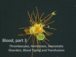
Blood, part 3 Thrombocytes, Hemostasis, Hemostatic Disorders, Blood Typing and Transfusions
Thrombocytes (platelets) • Not true cells • Cytoplasmic fragments w/purple granules that contain chemicals for blood clotting • Enzymes • Serotonin • Ca2+ ions • ADP • PDGF (Platelet-derived Growth Factor) • Normally kept in “inactive” state by molecules secreted by endothelial cells of blood vessels • When activated, form temporary plug that helps to seal breaks in blood vessels Activated platelet amorphous w/extensions that help them stick to vessels and each other
Thrombopoiesis (Platelet formation) • Cytoplasmic fragments derived from megakaryocytes • Mega = large • Karyo = nuclei • Cyte = mature cell • Thrombopoietin promotes formation of megakaryocyte, created by multiple mitosis w/o cytokinesis • Megakaryocyte presses against sinusoid (capillary) of bone marrow and pinches off to make fragments
Hemostasis • Series of reactions to stop a bleed • Phases: • Vascular spasm • Platelet plug • Coagulation (clotting) • Clot Retraction • Fibrinolysis (Clot Eradication)
1. Vascular Spasm 1. Vascular Spasm • Constriction of damaged vessel, triggered by: • Endothelium • platelets • - Pain receptors • Slows the bleed
2. Platelet Plug Formation • Damaged endothelium activate platelets by releasing • VWF (von Willebrand Factor) • Platelets become amorphous: • Stick to exposed collagen fibers of endothelium • Release • serotonin ( spasms) • ADP (attract more platelets) • Thromboxane ( both ) • Prostacyclin (inhibits aggregation elsewhere)
3. Coagulation (Blood Clotting) • Blood transformed from liquid to gel • 3 Critical Steps: • Prothrombin Activator Forms (complex series of biochemical rxns!) - require Ca2+ ion and procoagulants to promote sequence of rxns - Two pathways: a. Intrinsic Pathways (slower) – within damaged vessel b. Extrinsic Pathway (faster) – outer tissue around vessel 2. Prothrombin converted to Thrombin 3. Thrombin catalyzes joining of fibrinogen into fibrin mesh - Glue platelets together
4. Clot Retraction • Clot Retraction • Platelets contract (like a muscle) to stabilize clot • Contractions pulls on fibrin squeeze out serum from clot Compact the clot Ruptured ends of blood vessel drawn closer together • PDGF and VEGF released from platelets • (PDGF) Stimulates fibroblast and smooth muscle mitosis to rebuild wall • (VEGF) Promotes endothelial restoration
5. Fibrinolysis • tPA(Tissue Plasminogen Activator) • Released by endothelium around clot • Activates plasminogen • Plasminogen plasmin • Plasmin • Digestive “clot busting” enzyme • Remnants of clot phagocytized by WBCs • Macrophages • eosinophils
How are clots prevented from becoming TOO large? • Fast moving blood • Washes away excess Thrombin 2. Inhibition of clotting factors • Anti-thrombin III (ATIII): plasma protein inactivates thrombin • Protein C: inhibits clotting factors • Heparin: enhances ATIII
Hemostatic Disorders:Undesirable Clotting • Thrombus: a clot that develops in an unbroken vessel • blocks circulation • tissue death • Coronary thrombosis Heart attack Death of cardiac muscle
Hemostatic Disorders:Undesirable Clotting • Embolus: freely floating thrombus in bloodstream • Pulmonary Embolism • Impairs O2 delivery • Death of lung tissue • Cerebral Embolism • Stroke, • Death of brain tissue
Hemostatic Disorders:Undesirable Bleeding Thrombocytopenia • reduced platelet count < 50,000/mm3 • Bone marrow disease or destruction • petechiae on skin due to widespread hemorrhage • Treatment: blood transfusions
Hemostatic Disorders:Undesirable Bleeding Hemophilia: • Hereditary • Lack of clotting factors • hpA - defective Factor VIII (83%) • hp B - defective Factor IX (10%) • Genentech. Inc. - now engineers tPAand Factor VIII less transfusions needed
Human Blood Groups • glycoprotein antigens exist on external surfaces of RBCs • Antigens are: • Unique to the individual • “foreign” to other individuals • promotes agglutination • Presence or absence of these antigens is used to classify blood groups
Blood Typing • anti-A or anti-B antibody serums are added to blood • agglutination occurs between the antibody and the corresponding antigens
Rh Blood Groups • Rh antigens: • 8 types • Most common: types C, D, and E • Anti-Rh antibodies do not exist in Rh- individuals until they are exposed to Rh+blood (1st pregnancy) • Leads to complications during 2nd pregnancies, if Rh- mother is carrying Rh+ child
Transfusions • Blood bank: • Mix donor blood with anticoagulants that bind to Ca2+ • Tested for viruses, ABO, and Rh factor • 35 day shelf life at 40 C • Blood separated into component parts • Whole blood: (rare) • Only for rapid and substantial blood loss • Packed red cells (plasma removed) • To restore O2 carrying capacity
Transfusion Reactions • Occur when mismatched blood is infused • Donor’s blood cells are attacked by the recipient’s plasma antibodies causing: • Ruptured RBCs • Diminished O2 carrying capacity • Clumped cells (agglutination) • Blood clots • “Free” hemoglobin due to lysis of RBC • Hemoglobin solidifies in kidneys • Kidney fails