Fluoroscopy Credentialing
Fluoroscopy Credentialing. William Robeson, Radiation Safety Officer North Shore University Hospital Radiology/Radiation Safety Office (516) 562- 3895 Presentation created by: Miyuki Yoshida-Hay. Objectives.

Fluoroscopy Credentialing
E N D
Presentation Transcript
Fluoroscopy Credentialing William Robeson, Radiation Safety Officer North Shore University Hospital Radiology/Radiation Safety Office (516) 562- 3895 Presentation created by: Miyuki Yoshida-Hay
Objectives The Joint Commission accreditation requires physicians who use or operate fluoroscopic x-ray systems be properly credentialed. This training shall include radiation safety, management of fluoroscopic radiation and operation of fluoroscopic x-ray system(s) used by the physician. Training in radiation safety and fluoroscopic radiation management is in addition to any clinical training or qualifications required to perform the specific clinical diagnostic or therapeutic procedures for which the fluoroscopic systems are used.
Credentialing Requirements • The hospital requires documentation of appropriate training before granting fluoroscopic privileges. • In order to become credentialed, a physician must do the following: • View this powerpoint presentation • Complete the self-assessment quiz • Upon completing the training, each physician will need to complete an attestation. The attestation certificate is available for printing. • Present this certificate to the department chairperson for recording the successful completion of the fluoroscopy safety requirement. • It is up to the each department to establish other training and education requirements needed to obtain fluoroscopy privileges.
TOPICS • X-ray Production • Units of Radiation Exposure and Absorbed Dose • Dose Equivalent and Effective Dose Equivalent • Mobile C-arms • Factors Influencing Fluoroscopy Exposure Rate • Sources of Radiation Exposure • Radiation Protection • Personnel Monitoring • Basic Radiation Biology • Example of a Skin Injury from Fluoroscopy • Radiation and Pregnancy • Summary
X-ray Production X-rays are produced in an x-ray tubewhen electrons are accelerated through a high voltage (50,000 – 150,000 volts or 50 - 150 kVp) and allowed to hit a target composed of high atomic number materials such as tungsten.
X-ray Production (cont.) • Electrons are released from an electrically heated filament and are accelerated to the target by the high voltage. This flow of electrons from the filament to the target is known as the tube current (mA).Fluoroscopy is usually performed using 2 to 6 (mA) and an accelerating voltage of 75 to 125 kVp. • The amount of x-rays produced is determined by the tube current (mA) and the high voltage (kVp). X-ray production is directlyproportional to the tube current therefore doubling the tube current (mA) doubles the # of x-rays produced at a particular kVp. • However, x-ray production increasesmore rapidly with kVp than mA therefore increasing the kVp by 15% is equivalent to doubling the mA. NOTE: Higher kVp values also provides a more penetrating x-ray beam
Units of Radiation Exposure & Absorbed Dose • Exposure • The quantity of x-rays or gamma radiation required to produce an amount of ionization (electric charge) in air at standard temperature and pressure • Units: Roentgen (R) • 1R = 2.58x10⁻⁴ C/kg (coulombs/kilogram) • Usually expressed in terms of exposure rate: i.e., R/hr or fluoroscopy output measured as R/min. • Absorbed Dose • The amount of ionizing radiation energy absorbed per unit mass of tissue. • Units: rad (radiation absorbed dose) 1 rad = 0.01 Joules/kg 1 rad = 0.01 Gray • For x-rays used in fluoroscopy an exposure of 1R results in an absorbed dose of approximately 1 rad.
Dose Equivalent and Effective Dose Equivalent • Dose Equivalent • Used to account for differences in the biological effectiveness of different types of ionizing radiation. • Defined as (the absorbed dose) x (radiation quality factor) specific to the type of radiation to which an individual was exposed. • Units: rem (roentgen equivalent in man)or Sievert (Sv): 1 Sv = 100 rem • For diagnostic medical x-rays the quality factor is 1, therefore an absorbed dose of 1 rad is equal to a dose equivalent of 1 rem • Effective Dose Equivalent (EDE) • The risk of potential health effects when only part of the body is irradiated is smaller than when the whole body is exposed. • The EDE is a calculation of risk to an individual posed by a partial body irradiation. • The EDE is also used to estimate the equivalent whole body exposure for fluoroscopy staff wearing protective aprons for comparison to annual personnel dose limits for radiation exposure
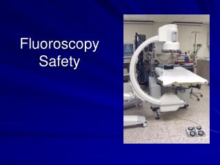
Mobile C-arms • Moved around ORs to visualize anatomy during surgery • Metallic c-arm contains x-ray tube at one end and image receptor at the other • An associated unit contains the display
Factors Influencing Fluoroscopy Exposure Rate • Modern fluoroscopy units produce images with an image intensifier (II) which brightens the image level sufficiently so that the image may be displayed on a TV screen. Fluoroscopy units are usually operated in an automatic brightness control (ABC) mode. • These units will automatically adjust the brightness by first increasing the kVp to increase x-ray penetration and then adjusts the mA to increase intensity. Note: Exposure to a thick patient will be greater than to a thin patient and also abdominal fluoro will require a greater exposure than a chest fluoro due to increased thickness and tissue density in the abdomen.
Factors Influencing Fluoroscopy Exposure Rate(cont.) Recommendation # 1: The image intensifier input should be positioned as close to the patient as practicable. This results in a lower patient dose and sharper image.
Factors Influencing Fluoroscopy Exposure Rate (cont.) Recommendation # 2: Use the exposure pedal as sparingly as possible. Radiation exposure during fluoroscopy is also directly proportional to the length of time the unit is activated by the foot pedal. Depression of the foot pedal determines the length of exposure. The fluoroscopy time is an important determinant of patient and staff radiation dose. Fluoroscopy units are equipped with a timer and an alarm which sounds at the end of 5 minutes. The alarm serves as a reminder of the elapsed time.
Factors Influencing Fluoroscopy Exposure Rate (cont.) Recommendation # 3: Use “last-image” hold and pulsed fluoro whenever possible. Most modern fluoro units are equipped with “last-image” hold, which stores the last fluoro image and allows viewing without having to expose the patient again. Many fluoro units also offer a “pulsed fluoro mode”, in which the x-ray beam is pulsed rapidly on and off and results in a lower radiation dose without significantly degrading the appearance of the image on the display.
Factors Influencing Fluoroscopy Exposure Rate (cont.) Recommendation # 4: Use the smallest field of view practicable. Radiation exposure also depends on x-ray field size and keeping the x-ray field as small as possible (by using collimators) which will decrease the dose to BOTH the patient and staff in the fluoroscopy suite. Restricting the field size not only decreases radiation dose but will also produce a better image. The contrast in the image between various tissue types will be greater for the smallest field of view that encompasses the desired anatomy.
Factors Influencing Fluoroscopy Exposure Rate (cont.) Recommendation # 5: High dose or detail modes should be used only sparingly. Many fluoro units will have various dose modes, such as low dose, medium dose and high dose mode. It is important to recognize that fluoroscopic image quality can be adversely affected by too few x-rays in the image; the image is noisy for low dose. More tissue contrast is produced by the “high dose” mode which will improve the image quality at the expense however of increased patient dose.
Factors Influencing Fluoroscopy Exposure Rate (cont.) Recommendation # 6: Magnification should be used only when necessary. Fluoroscopy units are capable of using different magnification modes. Image resolution is improved with magnification but field size is reduced and patient radiation dose is increased. Patient dose is minimized by using the lowest magnification (largest field size) appropriate for the image procedure being performed. Under Normal mode, there is little magnification with the whole beam used to generate a bright image. Under Mag 1 mode, a smaller beam area is projected to the same II output. The resulting object size is larger, but the image is dimmer due to the less beam input. The ABC system senses the brightness loss and either boosts machine X-ray output, increases tube voltage, or a combination of both.
6 inch mag FOV increases dose by a factor of 4 over non-mag image
Factors Influencing Fluoroscopy Exposure Rate (cont.) Recommendation # 7: For C-arm type fluoroscopy units the patient should be positioned as far from the x-ray tube as practicable to minimize patient entrance dose. To reduce personnel exposure the x-ray tube should be positioned beneath the patient.
In the case of portable C-Arm systems, eliminating the air gap between the I-I and the patient ensures that the table top is as far away as possible from the X-ray tube, minimizing radiation exposure to the patient’s skin. Note: The separator cone should always be utilized before commencing fluoroscopy on portable C-arm systems, as depicted below on the image at right.
Factors Influencing Fluoroscopy Exposure Rate (cont.) In conventional under-table x-ray tube fluoroscopic units, the x-ray tube is located at a fixed distance from the patient’s skin. In C-arm fluoroscopy, where the distance between the x-ray tube and image intensifier is fixed, the patient can be positioned in close proximity to the x-ray tube which increases the entrance skin dose and reduces image sharpness. It is preferable to locate the C-arm x-ray tube underneath the patient. Since the radiation transmitted through the patient is typically only 5 – 10 % of the entrance dose, inadvertent exposure to the operator hand on the exit side of the patient will result in a smaller dose compared to the dose to the hand on the entrance side of the patient. Also the amount of scatter radiation the operator is exposed to on the beam exit side of the patient is significantly less than on the beam entrance side.
X-ray tube X-ray tube Sorenson, 2000. Note: The benefit is exaggerated - some operator dose occurs on the image intensifier (I-I) side.
Sorenson, 2000. Care should be taken whenever the image view angle is changed during the procedure (e.g, changing from an ANT to a steep LAO). The I-I is often moved away from the patient while changing X-ray tube position. Large air gaps can result if the table or I-I height remains unadjusted.
Sources of Radiation Exposure • DIRECT EXPOSURE • Entrance Skin Exposure (ESE) rates (where the x-rays enter the patient) are limited to less than 10 R/minute(NOTE: At ESE rates of 10 R/min, 30 minutes of fluoroscopy can deliver 300 R in skin dose.) • ESE rates for typical fluoroscopy procedures are usually less than 5 R/min. • On some machines an operator can deliberately choose a setting that will increase the output. The use of higher radiation rates or "boost" modes are useful in situations requiring high video image resolution. ESE of up to 20 R/min is permitted for short duration. Special operator reminders, such as audible alarms, are activated during "boost" modes.
Sources of Radiation Exposure • SCATTER EXPOSURE TO PERSONNEL • Most of the radiation exposure received by the operator or other personnel in the fluoroscopy suite is due to scatter radiation from the patient. • The operator will be exposed to a dose rate of approximately one one-thousandth (1/1000) of the ESE rate at a distance of 1 meter from the center of the fluoroscopy field. Sorenson, 2000.
Sources of Radiation Exposure • Factors which increase the dose from scatter radiation: • Large patients which will cause the automatic brightness control (ABC) to adjust the kVp and mA to higher values causing greater amounts of scatter radiation. • A large x-ray field, a result of not restricting field size will increase scatter radiation • The length of time the fluoroscopy unit is on. Complex interventional cases will require greater procedure time, increasing dose to both the patient and operator
Sources of Radiation Exposure • Other sources of exposure to the operator may be associated with the following: • A small percentage of exposure to the operator may be due to leakage radiation through the x-ray tube housing. • C-arm operators should be aware that the shielding built in to “fixed” fluoroscopy systems is not available for protection against backscatter. This may be of greater concern if the C-arm is rotated out of the normal vertical plane.
Radiation Protection • The three most productive means of reducing radiation dose is: • Time: Minimize time spent in the radiation field. Use of “last-image-hold” and pulse fluoro features are technical advantages in reducing the total time x-rays are produced • Distance: Radiation dose rates increase or decreaseaccording to the inverse square lawEx: Double your distance from the source and decrease your exposure by a factor of 4 • Shielding: Use of lead garments, lead gloves, thyroid shields, leaded eyeglasses, lead drapes and clear leaded glass barriers between the patient and operator
Personnel Monitoring • Even when radiation protection techniques and engineering controls are in place to reduce personnel exposure, individual dose monitoring is required. • Various types of dosimeters are available (i.e., film badges, thermo-luminescent (TLD) and optically-stimulated luminescent (OSL) badges). • Badges are assigned to an individual and must never be shared. • A badge designed to measure the whole body (torso including head) should be worn at the collar – OUTSIDEthe lead apron.
Monthly Investigational Levels (mrems) • Level ILevel II • Body Badge (DDE) 50 150 • Collar Badge 150 450 • Eye (LDE) 150 450 • Ring/Wrist (SDE) or Extremity 500 1,500 • DDE = Deep Dose Equivalent • LDE = Lens Dose Equivalent • SDE = Shallow Dose Equivalent
Monthly Investigational Levels • Level I: Each incident will be noted on the personnel badge report by the Radiation Safety Office. A notification letter is sent to the employee. • Level II: The Radiation Safety Office will investigate each such incident. A report will be generated and the results of each investigation will be presented to the hospital radiation safety committee. • Physicians performing fluoroscopy that receive Level II collar badge readings will get a notification letter that includes their effective dose.
Personnel Monitoring (cont.) • The effective dose equivalent (EDE) may be calculated in the following manner: • A two-badge system (waist and collar badges) is used to calculate an individual’s EDE by taking into account the protective factor of the lead apron. In this situation, one badge is worn OUTSIDE the lead apron (collar) and a second badge is worn UNDERNEATH the lead apron (waist). The EDE is calculated as follows:EDE = [1.5 x (waist)] + [0.04 x (collar)] • A one-badge system (collar badge ONLY) is where one badge is worn on the OUTSIDE of the lead apron. The EDE is calculated as follows: EDE = [0.3 x (collar)]
Personnel Monitoring(cont.) • Dosimeters must be promptly turned in and exchanged each month to give accurate assessments. • Badge reports are reviewed by the Radiation Safety Office. Notification letters are sent to individuals who exceed monthly “Level I” exposure limits. Investigational letters are sent to those who exceed monthly “Level II” exposures limits. A written response to the letter is required, which includes an acknowledgement and an explanation of the Level II exposure, if known. The letter is to be returned to the Radiation Safety Office within one week of receipt. • Copies of badge reports (3 months) must be posted in each department for individuals to review. • Badges are susceptible to heat and moisture damage. Badges not in use should be stored in a cool, dry place, away from any sources of radiation. Do not take dosimeters home or travel on a plane with a dosimeter or wear a dosimeter during a medical radiological procedure.
OCCUPATIONAL DOSE LIMITS (NRC) For hospital radiation workers, annual doses rarely exceed 10 % of these values.
Basic Radiation Biology • X-rays from fluoroscopy interact with biological materials by transferring their energy to an electron which subsequently interacts with the target molecule to produce an ion or a free radical. • Indirect action is the creation of free radicals from interactions with water molecules. Free radicals may then chemically interact with biologically sensitive molecules (DNA, RNA, proteins) causing damage • Direct action is the interaction of ionizing radiation with biologically sensitive molecules such as DNA causing direct destruction or mutation • Since water molecules are much more numerous than biologically sensitive molecules, indirect action is the most common form of biological damage.
Basic Radiation Biology (cont.) • Cells can sustain a variable amount of radiation and still repair themselves from sub-lethal damage. • Continuous high intensity radiation will produce greater damage than an equivalent fractionated (multiple smaller) dose since fractionation allows for cell repair.
Basic Radiation Biology (cont.) • A given organ’s response to radiation depends on: • Total dose • Dose rate • Fractionated scheme • Volume of irradiated tissue • Inherent tissue radiation sensitivity • A large total dose, high dose rate and small fractionated schedule (which is all possible in fluoroscopy) will cause a greater degree of damage. • Major concern in fluoroscopy is the possibility of acute, direct or deterministic, radiation damage which manifests as a skin injury. The severity of skin injury is dose-dependent; more dose means more severe symptoms
Note that the time to expression of symptoms is long enough that the patient may no longer be in the hospital when symptoms appear. The physician performing the fluoroscopy cannot discern the damage by observing the patient immediately following the procedure.
Basic Radiation Biology (cont.) These threshold doses to cause an effect cannot be considered exact due to many variables such as individual biological response, age, characteristics of the individual exposed and the area exposed. A patient may exceed the threshold dose without showing symptoms. This may be due to: The x-ray beam may not have been concentrated on a single area of the skin for the entire time and because 10 R/min or 20 R/min are maximum outputs for very thick patients.
Example of a Skin Injury from Fluoroscopy This case, patient A is that of a 40-year-old male who underwent coronary angiography, coronary angioplasty and a second angiography procedure due to complications, followed by a coronary artery by-pass graft, all on March 29, 1990. The area of injury six to eight weeks following the procedures. The injury was described as "turning red about one month after the procedure and peeling a week later."
Example of a Skin Injury from Fluoroscopy (cont.) In mid-May 1990, it had the appearance of a second-degree burn. The condition in late summer 1990, exact date unknown, with the appearance of a healed burn, except for a small ulcerated area present near the center.
Example of a Skin Injury from Fluoroscopy (cont.) Skin breakdown continued over the following months with progressive necrosis.
Example of a Skin Injury from Fluoroscopy (cont.) The injury eventually required a skin graft. The magnitude of the skin dose received by this patient is not known. However, from the nature of the injury, it is probable that the dose exceeded 20 Gy. Wagner LK, Eifel PJ, Geise RA. Potential Biological Effects Following High X-ray Dose Interventional Procedures. JVIR 1994; 5:71-8
Deterministic Effects • Threshold dose below which no effect is observed • Severity increases with dose • Examples: skin erythema, dermatitis, desquamation cataracts
Stochastic Effects • Incidence increases with dose • No dose threshold assumed • Basis for ALARA principle of radiation protection • Example: cancer
Pregnancy and Radiation • The decision to perform a radiological procedure on a patient who may be pregnant is a medical decision and shall be made by a physician in consultation with the patient. If the procedure is to be performed, the physician must explain the risks to the patient, provide informed consent and the appropriate consent form shall be signed. • Shielding shall be used to shield the abdomen from radiation provided it does not interfere with the procedure. • Every attempt must be made to minimize direct exposure to the fetus according to the principles described in this presentation. • Medical emergency radiological procedures however take precedence over pregnancy status.
Principles for Fluoroscopy • PAUSE to properly plan and prepare for study • Activatedose saving features of equipment • Noexposures unless necessary • Depresslast image hold and last image grab instead • PULSE at lowest possible rate