Download
1 / 38
400 likes | 748 Views
Physician Credentialing For the Use of Fluoroscopy. Kim James RT-R, CV,CT, QA Coordinator CaroMont Imaging Services Edited by Jennifer Helton, RT-R, 2013. Preface. The following guidelines will aid physicians in minimizing the use of ionizing radiation in fluoroscopically guided procedures.

E N D
Physician Credentialing For the Use of Fluoroscopy Kim James RT-R, CV,CT, QA Coordinator CaroMont Imaging Services Edited by Jennifer Helton, RT-R, 2013
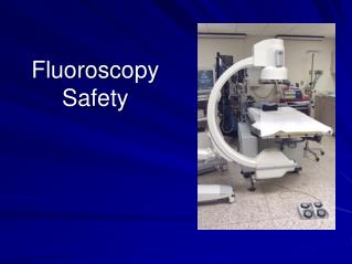
Preface • The following guidelines will aid physicians in minimizing the use of ionizing radiation in fluoroscopically guided procedures. • Physicians should be aware of the potential for serious radiation induced skin injury. • The following tutorial will advise you in the techniques to optimize the use of the C-arm and radiation protection practices thereby reducing the radiation dose to the patient, staff and yourself. • It is the physician’s obligation to protect the staff and the patient from over exposure to radiation.
Preface The North Carolina Radiation Protection Commission statue .0603 General Requirements (B) states ”Individuals who will be operating the x-ray equipment shall be instructed on the safe operating procedures and use of equipment and demonstrate an understanding there of.”
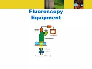
Definitions • ABC: (Automatic brightness control); The ABC compensates brightness loss caused by decreased image intensifier radiation reception by generating more X-rays (increasing mA) and/or producing more penetrating X-rays (increasing kVp). Conversely, when the image is too bright, the ABC compensates by reducing mA and decreasing kVp. • Interpolation: Sometimes called re-sampling is an imaging method to increase (or decrease) the number of pixels in a digital image. • Air Kerma: Is another radiation quantity that is sometimes used to express the radiation concentration delivered to a point, such as the entrance surface of a patient's body. • DAP: (Dose Area Product); The dose-area product is a measurement of the amount of radiation that the patient absorbs.
Definitions Inverse square law: The intensity or dose of the radiation emitted from the source of the X-ray beam diminishes with the square of its distance from the source. If you double the distance, the dose decreases to a factor of ¼. For example, if you received 8rads of radiation at 2ft., you would only receive 2rads of radiation at 4ft. MPD: Maximum Permissible Dose
ALARA • ALARA concept: • ALARA stands for As Low As Reasonably Achievable. • Because some risk, however small, exists from any radiation dose, all doses should be kept ALARA. • Includes reducing both internal and external radiation dose. • ALARA is the responsibility of all personnel involved in radiographic procedures.
Equipment Quality Control • When new equipment is installed it must be tested by a medical physicist. The equipment must meet FDA and state regulatory requirements, manufacturers specifications, and lowest radiation dose level settings to produce image quality. • Fluoroscopic equipment must be maintained in good working order with all electrical and radiation safety features within regulatory compliance. Annual inspections are performed by a medical physicist to ensure radiation safety and image quality. • Preventive maintenance is performed on an annual basis by a qualified service engineer. Tests will include proper calibration of the x-ray tube and the x-ray generator.
Units of Radiation Radiation is measured in units: • Roentgen - unit of radiation exposure in air • Rad - energy absorbed per gram of tissue • Rem - biological effect of a rad
Known Biological Effects of Radiation • Marrow depression 100 Rads • Cataracts 200 Rads • Erythema 200 Rads • Temporary reduced fertility 250 Rads in males 150 Rads in females • Permanent sterility 500 Rads in males 600 Rads in females • Birth defects in human fetus 10 Rads in first trimester
Exposure Limits • All persons who are associated with the operation of an x-ray system are subject to the occupational exposure limits. • The annual limit is the total effective dose equivalent, which is equal to 5 rems .
Exposure Limits MPD (mrem/yr) ALARA
Protective Clothing, Equipment and Monitoring • Lead Aprons: Must be worn by physicians and ancillary staff in a room where fluoroscopy is performed. The myth that a lead apron is worn to stop radiation is false, the truth being that it can only reduce the amount of radiation exposure to an acceptable level.Radiation loses energy and intensity as it passes through lead and will most likely be absorbed in the body tissue. • Thyroid shields and leaded glasses: added protection against cataracts and thyroid problems. • Personal dosimeters: collar and waist badges must be worn during fluoroscopic procedures to measure occupational exposure • Control badges
Protective Clothing and Equipment • RADPADS may be placed on the patient before creating a sterile field. RADPAD shields and drapes reduce harmful scatter radiation by as much as 95%. Sterile and disposable RADPAD Shields are effective in reducing scatter radiation in medical intervention utilizing x-radiation. • Sterile, lead-free and repositionable, RADPAD shields are placed directly on the patient to provide the physician or technologist with moveable protective device in which to work.
Cardinal Principles of Radiation Safety • Time • The less time spent near the radiation source, the less radiation received. • Distance • Inverse Square Law = double the distance, 1/4 the dose • Shielding • -Aprons: 0.25 – 0.5 mm lead equivalent, • -Thyroid collar, lead glasses, lead eye protection • -Ceiling mounted, rollaway shields, and RADPADS
The Most Common Sense Principle DISTANCE - One step back from tableside: cuts exposure by factor of 4 - Lateral fluoroscopy: 5x less dose on Image Intensifier side - Moving Image Intensifier closer to patient: the patient skin exposure is lessened there is less scatter radiation creating a sharper image - Source to Skin Distance (SSD): 38 cm for stationary fluoroscopes 30 cm for C-arm fluoroscopes - Distance is the most effective means of radiation protection.
Patient dose is directly proportional to the time your foot remains pressed on the fluoro pedal. Radiation Exposure Reduction
Best Radiation Exposure Reduction Practices • Distance from source • Collimation • Automatic Brightness control • Pulse Rate Reduction • Tube Angulation • Last Image Hold • Skin Entrance Dose Monitoring
Patient moved closer to x-ray tube. 30cm 60cm 2 R/min 8 R/min 60cm 30cm Exposure rate quadruples when the patient to focal spot distance is halved! The inverse square law • The Xray tube should be as far away from the patient as possible and the image intensifier as close to the patient as possible. “Ideal” setup
reduced x-ray field size collimators typical collimator control buttons on the control panel collimated x-ray beam uncollimated x-ray beam Collimation Restrict the x-ray field of view by using collimation. “Cone down” to the area of interest. Avoid irradiation of irrelevant anatomy, this reduces the volume of tissue irradiated, and reduces x-ray scatter, it also improves image quality by reducing scatter.
Automatic Brightness Control • Use the settings which give the lowest patient entrance exposure rates consistent with acceptable image quality. • Choose high kV / low mA options when available • “Record” modes such as cine, DA, and DSA produce the highest quality (lowest noise) images. **CAUTION!: Use high dose rate or “boost” fluoro modes as infrequently as possible. These double patient entrance exposure rates and are designed for situations when normal fluoro images are too noisy to properly carry out a procedure. They are almost never needed. It is a legal requirement that a continuous audible beeping signal be heard whenever the fluoro pedal is depressed in high dose or boost mode. .
· Although a smaller volume of tissue is irradiated in MAG mode, the radiation intensity roughly doubles with each step up in magnification. MAG modeRelative Exposure Rates Normal 1 MAG1 2 x normal MAG2 4 x normal Magnification Modes Use electronic magnification (MAG) modes sparingly. • MAG modes allow for the magnification of small areas for better visualization.
mA mA time time Pulse rate reduction For pulsed fluoro units, use the lowest pulse rate consistent with the resolution requirements of the procedure during cine and DSA applications. Lower Pt Dose Insertionof guide wires and similar procedures can usually be performed at low pulse rates - 15 or 7.5 pps (pulses per second). Even lower rates can be used if fluoro image interpolation is available or the physician has a high tolerance for jerky images and flicker.
Image still present Foot off pedal Last image hold C-arms at Caromont Regional come with a last image hold feature. The last image that was taken will remain on the TV monitor after the release of the fluoro pedal. This allows the fluoroscopist to view the image so that decisions and discussions about the image can occur with the radiation off.
Even small changes in tube angle can spread the dose over a wider area due to the curvature of the body. Variation of x-ray tube angulation When long fluoro times are anticipated (greater than approximately 15 min of actual x-ray beam “on” time) the tube angle should be changed so that the same area of skin is not under constant irradiation. · Always bear in mind that larger patients always receive higher dose rates.
Patient and Tube Positioning To reduce the chance of the patient receiving a significant skin dose, the patient should be placed as close to the image intensifier as possible. The x-ray tube should be as far away as possible from the patient. Ideal Can be dangerous due as a result of the inverse square law.
Dose-area product The dose-area product is a measurement of the amount of radiation that the patient absorbs. • The dose-area product is independent of the distance between the X-ray tube and the measuring device because the further away from the X-ray tube this measurement is taken, the more the size of the device increases, and the dose itself decreases. • The dose to the patient can be calculated from the dose-area product, the size of the measuring device, and the distance to the X-ray tube and the patient
Patient Skin Entrance Dose If the cumulative air kerma for a given case exceeds 1 Gy (1,000mGy), a qualified medical physicist needs to be called in to determine if the actual skin entrance dose exceeded the 1 Gy(100 rad)= regulatory threshold.
Fluoroscopy Skin Entrance Dose Monitoring • Effect of Different Fluoro Modes • a. Low dose level fluoro if available, will decrease the dose rate by 50% -100% with a commensurate increase in time before reaching 1 Gy. • b. Variable frame rates for pulsed fluoroscopy will generally result in a 50% reduction in dose rate going from 30 pps to 15 pps, and a 30% reduction from 15 pps to 7.5 pps with commensurate increases in time. • c. High dose rate or boost fluoro (audible continuous signal heard when the fluoro pedal is depressed) - time before reaching threshold will be halved.
Fluoroscopy Skin Entrance Dose Monitoring • DSA ‘Record’, Cine, or DA modes of operation increase exposure rates by factors of 10 or greater and are highly dependent upon the x-ray unit itself, the frame rate, MAG mode, technique, and dose rate level selected. • Example: For a typical cardiac angio system with a cine patient entrance exposure rate of 40 R/min (medium to large patient), the 100 rad threshold would be reached in 2.5 min for a PA projection. For a large angle oblique, the dose rate could double or triple and the time reduced to approximately 1 min.
Larger area - less intense field DAP’ = K’A’ DAP = K A DAP’ = DAP Smaller area - more intense field DAP does not depend on distance Fluoroscopy Skin Entrance Dose Monitoring • Dose Area Product (DAP): Air kerma x X-ray field area. If you know the field size, A, at the patient’s skin you can estimate the skin entrance dose: K = DAP / A K= Air Kerma DAP= Dose Area Product A= x-ray field area • An ion chamber located at the exit of the x-ray tube collimator assembly • directly measures the air kerma exiting the tube. • · Sensors measure the size of the x-ray field at this position. • · A microprocessor calculates the product: DAP = KA for display
An ion chamber located at the exit port of the x-ray tube collimator assembly directly measures the air kerma exiting the tube. Fluoroscopy Skin Entrance Dose Monitoring Patient Air Kerma Readout: All fluoroscopic units manufactured after June 10, 2006 must comeequipped with air kerma rate (AKR) and cumulative air kerma (AK) displays on the monitor the patient skin entrance dose during each procedure.
Fluoroscopy Skin Entrance Dose Monitoring The table below gives ballpark estimates of exposure times required to reach 1 Gy (100 rad) skin entrance dose.
Pregnant patients There is little that can be done to reduce fetal doses other than practicing the radiation reduction techniques and careful collimation. • When the fetus is outside the direct x-ray field, the fetal dose is caused almost entirely by internal scatter from the mother. This cannot be avoided. • Since the amount of scatter is proportional to the volume of tissue directly irradiated, • Collimating down reduces the fetal dose proportionately. fetus
Pregnant patients The ABC feature will dramatically increase the kV /mA if the apron partially blocks one of the light sensors - overdosing adjacent tissue in the x-ray field, increasing scatter, and dramatically reducing image quality (glare).
Pregnant patients The draping of lead aprons on the mother, posterior and/or anterior to fetus, will have a minimal impact on the fetal dose. • However,If the fetus is very close to the edge of the x-ray field, a lead apron will protect it from inadvertent direct radiation. Furthermore, the use of lead often has a comforting affect on the mother (and family) and may be used for this purpose. fetus
Pregnant patients If the fetus is unavoidably in the direct x-ray beam (and is not the object of interest), using a lead apron to partially block the x-ray beam is generally not recommended.
Documentation It is required by state and federal laws to maintain a log at each fluoro station and C-Arm stating the following information: • Patient Name • Patient Identifier ( Medical Record Number) • The Name of Procedure • The Operator (Physician Name) • The Fluoro Time