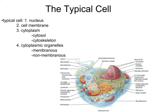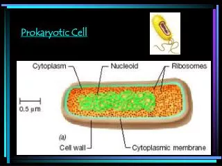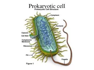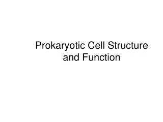
Typical Prokaryotic Cell
Typical Prokaryotic Cell. Prokaryotic Cell Structures. Functions of Cell Membrane. 1. Selective barrier (selectively permeable) 2. Secretes exoenzymes amylases lipases peptidases. Functions of Cell Membrane. 3. E.T.S. is located here 4. Enzymes for cell wall synthesis
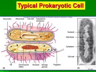

Typical Prokaryotic Cell
E N D
Presentation Transcript
Functions of Cell Membrane • 1. Selective barrier (selectively permeable) • 2. Secretes exoenzymes • amylases • lipases • peptidases
Functions of Cell Membrane • 3. E.T.S. is located here • 4. Enzymes for cell wall synthesis • 5. If photosynthesis, enzymes are located on membranous structures called thylakoids • 6. Mesosomes - invagination of cell membrane attached to DNA (Binary Fission)?
Cytoplasmic Membrane • Movement across membrane for many substances is controlled by membrane proteins. • Escherichia coli has >200 membrane proteins. • Many of these proteins are involved in transport across membranes. • Others of these proteins allow a bacterium to sense its surrounding environments (e.g., as in chemotaxis). • Movement is via: • Simple Diffusion (including osmosis) • Facilitated Diffusion (with concentration gradient & no energy expended) • Active Transport (against concentration gradient & energy expended)
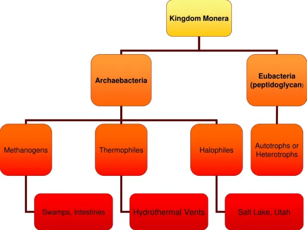
The Prokaryotic Cell Wall In some cases recognized by host immune system. Determines cell shape. Prevents osmotic lysis. Target for antibiotics. In Bacteria, composed of Peptidoglycan. Part of cell envelope.
Cell Wall • Main structural component - Peptidoglycan • Peptidoglycan • repeating dissacharide units • polypeptides
Gram-Negative Cell Envelope endotoxin cell wall
Gram-Negative Cell Envelope LPS: Protection from antibiotics such as penicillin plus against certain toxins. Periplasm: Site of preliminary nutrient degradation.
Lipopolysaccharide (LPS) Carbohydrate has negative charge and provides protection against some antibiotics & some toxins (e.g., detergents). Lipid A = Endotoxin
Mycoplasma lack Cell Walls Note: Pleomorphic Mycoplasma pneumoniae causes “Walking Pneumonia”
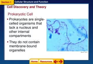
Prokaryotic Cell Structure • Glycocalyx - term to describe substances that surround bacterial cells • 1. Capsule • if substance is organized and firmly attached to cell wall • 2. Slime Layer • if substance is unorganized and loosely attached to cell wall
Function of Capsule 1. Contribute to Virulence of bacteria by preventing phagocytosis by WBC’s A. Streptococcus pneumoniae B. Bacillus anthracis
Functions of Capsules • 2. Prevents drying out or dessication • 3. Allows bacteria to adhere to various surfaces • Streptococcus mutans - enamel on teeth to cause dental carries • Klebseilla pneumoniae - attaches to respiratory tract
Glycocalyx Protection(e.g., Streptococcus pneumoniae from phagocytosis) Attachment(e.g., Streptococcus mutans causing dental plaques)
Capsule Staining Capsules are more regular and gelatinous. Slime Layers are less regular and more diffuse.
Flagellar Arrangements Polar Flagellum e.g., E. coli also “atrichous”
Chemotaxis Also Phototaxis, etc.
Fimbriae (a kind of pilli) Tips are Adhesins, used to adhere, e.g., to animal tissues
Motility • Almost all Spiralbacteria aremotile • About 1/2 ofBacilliaremotile • Almost all Cocciare non-motile
Axial Filament - found only in spirochetes (flexible spirals) Treponema pallidum
Fimbriae • Filamentous appendages that are shorter, straighter and more numerous that flagella • found mostly in Gram (-) Bacteria • used for attachment not motility
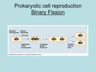
Nuclear area (nucleoid) • 1 circular chromosome (ccDNA) • attached to a mesosome • segragation of DNA during Binary Fission
Plasmids • Small circular, extra-chromosomal pieces of DNA • 5 to 100 genes • Code for auxiliary metabolic functions: • antibiotic resistance • penicillase • production of toxins • E. coli 0157:H7
Prokaryotic Ribosome 70 S 50 S 30 S Eukaryotic Ribosomes 80 S 60 S 40 S Ribosomes - protein synthesis
Selective Toxicity • Some antibiotics are aimed at the 70 S ribosomes of bacterial cells • Streptomycin, Neomycin, Erythromycin and Tetracycline work by inhibiting protein synthesis by disrupting the 70 S ribosome
Endospores - formed under periods of environmental stress • Only found in Gram (+) Bacteria • Bacillus • Bacillus cereus • Bacillus anthracis • Clostridium • Clostridium tetani • Clostridium botulinum • Clostridium perfringens
Endospores • Extremely resistant to heat, cold, chemicals, lack of water, etc. • Most vegetative bacterial cells are killed at temps. above 70 C (160 F) • Endospores can survive boiling water for several hours (some for as long as 20 hours)
Endospores • Spores can remain viable for weeks, months, years • Thermoactinomyces vulgaris • spores found in Minnesota were 7,500 years old and still germinated
Form inside of vegetative cells (hence “endo”). Endospores Characteristic of many soil bacteria, e.g., Bacillus spp. & Clostridium spp. Highly resistant to heat, U.V., desiccation, etc.