Download
1 / 23
250 likes | 616 Views
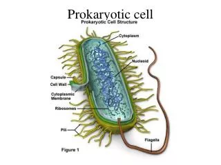
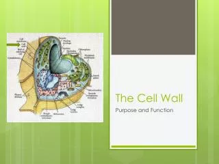
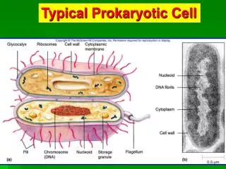
The Prokaryotic Cell Wall. Functions of the Cell Wall. Shape provided Protection for osmotic shock (lysis) If water moves in lysis (in a hypotonic environment) If water moves out plasmolysis (shriveling) (in a hypertonic environment) Contribute to pathogenicity (e.g. LPS)

E N D
Functions of the Cell Wall • Shape provided • Protection for osmotic shock (lysis) • If water moves in lysis (in a hypotonic environment) • If water moves out plasmolysis (shriveling) (in a hypertonic environment) • Contribute to pathogenicity (e.g. LPS) • Protection from toxic compounds
The cell wall can be removed experimentally • Lysozyme – hydrolyzes PTG • Penicillin – inhibits PTG syntesis • Result: • Gram-positive protoplasts • Gram-negative spheroplasts (OM intact) • Osmotically sensitive must be maintained in an isotonic solution
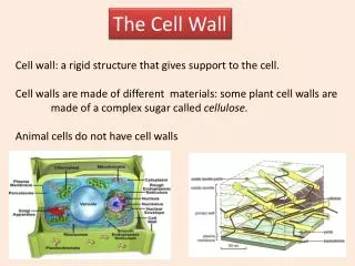
Differences between Gram-positive and Gram-negative Bacteria • Gram-positive cell wall • Composed of THICK PTG (murein layer) • Single layer – 20-80 nm • Located outside of PM • Contains large amounts of teichoic acid
Gram-negative cell walll • THIN PTG • 1-3nm • Surrounded by an outer-membrane located outside of PM • Contains lipopolysaccharide (LPS = endotoxin)
Periplasmic Space • A gap between outer-membrane and plasma membrane of Gram-negative bacteria • A gap between cell wall and plasma membrane of Gram-positive bacteria • Filled with periplasm • Proteins used for metabolism, protection and export
Peptidoglycan Structure • Large polymer made up of alternating subunits of: • N-acetylglucosamine (NAG) • N-acetylmuramic acid (NAM) • Unique to prokaryotic cells
Tetrapeptide attached to NAM at carboxyl group • Alternating D- and L- amino acids • L-alanine • D-glutamic acid • Meso-diaminopimelic acid (L-lysine in Gram-positive bacteria • D-alanine • (D-amino acids and meso-diaminopimelic acid not found in proteins)
Gram-Positive Strategy • Polymeric chains of PTG stabilized by cross-connecting bridges of amino acids in Gram-positive bacteria • Cross-links form an interbridge • Pentaglycine peptide bridge utilized • Covalently link D-ala (4th amino acid) of tetrapeptide attached to NAM with L-lysine (3rd amino acid) of another tetrapeptide • Strengthens cell wall – Dense network - THICK
Gram-Negative Strategy • Carboxyl group of D-ala (#4) is covalently linked too amino group of diaminopimelic acid (#3) • No pentaglycine bridge is involved • Tetrapeptides themselves are the bridge • Often have “free” tetrapeptides • Lipoproteins sometimes bound to NAM • Thinner than Gram-positive PTG components
Gram-positive cell walls • 20-80nm thick • 60-80% extensively cross-linked PTG • Forms a thick polymeric mesh • Peptide bridge can vary in length and composition (i.e. not always pentaglycine)
Often contain teichoic acids • Linked to muramic acid of PTG via phosphodiester bonds • Two major forms • Ribitol teichoic acid • Long polymer of ribitol • 5 carbon sugar alcohol • Glycerol teichoic acid • Long polymer of glycerol • 3 carbon sugar alcohol
Sometimes linked to PM lipids (lipoteichoic acids) • Extend through peptidoglycan layer to external surface • Highly antigenic • Antibodies often used for serological identification of different strains of Gram-positive bacteria
Gram-negative bacteria • More complex chemically than Gram-positive cell walls • Less PTG (only 5-10%, much thinner – 1-3nm, usually a monolayer) • Second membrane structure located exterior to PTG = Outer-membrane (OM) • Composed of protein, phospholipid, lipoprotein and lipopolysaccharide (LPS) • Braun’s lipoprotein – anchors OM to PTG • LPS = toxic to animals • AKA endotoxin • Causes high fever, tissue injury, cell death and shock • Macrophages contain LPS-binding receptors induces secretioon of TNF-alpha and IL-1
LPS - Three Major Components • Lipid A • Two glucosamine sugar derivatives inked to fatty acids • Phosphate or pyrophosphate attached • Responsible for toxicity • Buried in OM • Core polysaccharide • Linked to Lipid A • Often contains unusual sugars (e.g. heptuose) • Contains phosphate • O antigen • AKA somatic antigen • Short polysaccharide often with unusual sugars (e.g. mannose, galactose) • Different types depending on strain • Antibody responses directed towards this antigen (useful for serotyping)
Functions of LPS • Stabilization of OM • O side chain composition can be changed to avoid host antibody detection • Prove net negative charge on the cell
Function of OM • Protective barrier • Inhibits or regulates toxic compounds, metabolites • Permits entry of small molecules through porin proteins • Prevents loss of periplasmic enzymes
Gram-positive and Gram-negative bacteria stain differently because of the composition of their cell walls • PTG layer retains crystal violet more efficiently in Gram-positive bacteria because of its thickness and degree of cross-linking (smaller pores) • Crystal violet/iodine complex more easily removed from ram-negative bacteria
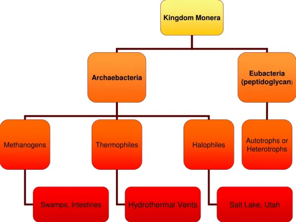
What makes Archaebacteria unique? • Archaebacteria (unlike eubacteria) do NOT contain PTG • Possess unusual lipids in their membranes • Branched chain hydrocarbons connected to glycerol by ether links • May also contain • Pseudomurein • Polysaccharides • Glycoprotens • Differences in tRNA and RNA polymerases structure • Diverged evolutionarily from eubacteria before the origin of eukaryotes
What makes Mycoplasma unique? • Mycoplasma lack a cell wall • They use sterols for strength
What makes L forms unique? • L forms lack cell walls • They arise by spontaneous mutations or when grown in isotonic medium containing penicillin