
Cardiovascular Disease
E N D
Presentation Transcript
Cardiovascular Disease J. Matthew Velkey matt.velkey@duke.edu 454A Davison, Duke South, Green Zone
Intimal thickening • Endothelial injury: • mechanical: hypertension, turbulent flow, catheterization • inflammatory: stasis, infection, auto-immune response • toxic: oxidized lipids, cigarette smoke • Vascular smooth muscle cell recruitment and proliferation • Elaboration of extracellular matrix
ARTERIO-SCLEROSIS • GENERIC term for ANYTHING which HARDENS arteries • Atherosclerosis (99%) • Mönckeberg medial calcific sclerosis (1%) • Arteriolosclerosis, involving small arteries and arterioles, generally regarded as NOT strictly being part of atherosclerosis, but more related to hypertension and/or diabetes
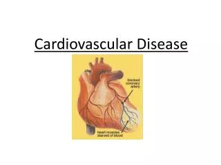
ATHEROSCLEROSIS • Chronic endothelial injury • LDL, Cholesterol in arterial WALL • OXIDATION of lipoproteins • Monocytes migrate endothelium* • Platelet adhesion and activation • Migration of SMOOTH MUSCLE from media to intima to activate macrophages (foam cells) • Proliferation of SMOOTH MUSCLE and ECM • Accumulation of lipids in cells and ECM (fatty streak) • Formation of PLAQUES
Four major players in atherosclerosis 1) Endothelial Injury 2) Inflammation 3) Lipids 4) Smooth Muscle Cells, SMCs
Progression of Atherosclerotic Plaques Fatty streak ↔ Fibrofatty Plaque Vulnerable Plaque Once a vulnerable plaque: aneurysm OR rupture and thrombosis OR critical stenosis
FATTY STREAKS
Atherosclerotic plaques • Fibrous cap: VSMC, macrophages, foam cells, lymphocytes, extracellular matrix • Necrotic center: cell debris, cholesterol clefts, foam cells, calcification
Microscopic features Lumen, Fibrous cap (fibrous plaque), Lipid core, External Elastic Membrane thinning/destruction, Calcification, Neovascularization
Hypertension • Sustained systolic > 140 or diastolic > 90 • Pre-hypertension: systolic between 120-139, diastolic between 80-89 • Often (95%) multi-factorial etiology or “essential” • Less frequently (5%) known to be “secondary” to another condition Diuretics ACE inhibitors β blockers α blockers
HISTOPATHOLOGY ofESSENTIAL HYPERTENSION “hyaline” = benign HTN “hyperplastic” = malignant HTN, systolic>200 1) onion skin 2) “fibrinoid” necrosis
Aneurysms and dissections • TRUE: Weakness in vessel wall causes it to bulge symmetrically (fusiform) or, more often, asymmetrically (saccular) • FALSE: hemorrhage and formation of a hematoma may give the appearance of a saccular aneurysm • DISSECTION: tear in intima causes separation along intima-media junction
Aneurysms 2 CAUSES: • ATHEROSCLEROSIS • Cystic medial degeneration (e.g. Marfan syndrome, Ehlers-Danlos syndrome) Normal aorta Aorta with medial degeneration
Most abdominal aortic aneurysms (AAA) occur between the renal arteries and the bifurcation of the aorta
Aneurysm Complications • RUPTURE or DISSECTION • OBSTRUCTION • EMBOLISM • COMPRESSION • Ureter • Spine • Trachea • Esophagus
Dissection • Separation of vessel wall (usually aorta and usually along intima-media junction. • Associated with aneurysm, hypertension, or trauma • “Type A” more common • Risk of rupture and massive internal bleeding • Also can constrict tributaries causing ischemia • Symptoms: severe sternal or interscapular pain, progresses downward with spread of dissection
VASCULITIS • Chiefly arterial • Infectious (5%) vs. Non-infectious (95%), often auto-immune • Often DRUG related (Hypersensitivity, e.g.)
“TEMPORAL” ARTERITISaka, Giant Cell Arteritis, GCA • ADULTS • Mainly arteries of the head and temporal arteries are the most visibly, palpably, and surgically accessible • BLINDNESS most feared sequelae • GRANULOMATOUS WALL inflammation diagnostic • Anti-NEUTROPHIL AB’s often POSITIVE
Raynaud “Phenomenon” • PRIMARY: exaggerated vasomotor response • Proximal vasodilation mid-digital vasoconstriction end digital cyanosis (WHITE) (BLUE) (RED) • Vasoconstriction usually triggered by COLD, emotion • Self-limited, ischemia & gangrene UN-common • Arteries often do NOT show diagnostic pathology other than some intimal thickening • SECONDARY: vascular insufficiency due to some other disease • Atherosclerosis, SLE, Buerger Disease, etc.
“Varicose” Veins • 20% of population, F>M • Related to increased venous pressure, age, valve dysfunction • Superficial veins of lower extremities most common, also external anorectal venous plexus (hemorrhoids) • If severe can result in ischemia (poor venous return) and ulceration • Can also occur in the setting of LIVER CIRRHOSIS –blood backs up within portal system and varicosities develop at porto-caval anastmoses (esophagus, internal rectal plexus, and abdominal wall) • rupture of esophageal varices can cause life-threatening internal bleeding
THROMBOPHLEBITIS(aka PHLEBOTHROMBOSIS) • 90% DEEP veins of the legs • Factors: • CHF (stasis) • Neoplasia (esp. GI, pancreatic, and Lung adenocarcinomas) often cause hypercoagulability • Pregnancy, obesity, post-op, immobilization, or any of the parts of Virchow’s triangle • Sequelae: PE most feared • Symptoms: edema, cyanosis, heat, pain, tenderness, but usually……..NONE!!!
SVC Syndrome • Blockage within SVC –backup causes decrease in arterial flow regions drained by SVC (can also involve pulmonary vessels) • Usually from bronchogenic carcinoma or mediastinal lymphoma • “DUSKY CYANOSIS” of: • Head • Neck • Arms
IVC Syndrome • Secondary to: • NEOPLASMS (external compression) • ASCENDING THROMBOSIS from FEMORALS, ILIACS • AAA, Gravid uterus • Bilateral leg edema • Massive proteinuria if renal veins involved (like nephrotic syndrome) • If hepatic veins involved, can cause liver congestion and necrosis
ISCHEMIC HEART DISEASE ISCHEMIC HEART DISEASE (IHD) CORONARY HEART DISEASE (CHD) CORONARY ARTERY DISEASE (CAD) ATHEROSCLEROTIC HEART DISEASE (ASHD) Synonymous terms referring to syndromes resulting in and from myocardial ischemia
Percentage Breakdown of Deaths From Cardiovascular Diseases United States:2003* Source: CDC/NCHS and NHLBI. *Preliminary
ISCHEMIC HEART DISEASE The underlying cause of ischemic heart disease is usually atherosclerosis of the coronary arteries The most common cause of acute coronary syndromes (unstable angina or acute myocardial infarction) is a sudden increase in luminal narrowing due to thrombosis and/or plaque rupture.
MAJOR SYNDROMES ANGINA PECTORIS “STABLE” (pain upon exertion) “UNSTABLE” (pain upon little to no exertion) Chronic Ischemic Heart Disease Myocardial infarct /sudden cardiac death
PREVALENCE OF ISCHEMIC HEART DISEASE 13.5 million Americans (7% of adult population) have symptomatic IHD evidenced by: Angina Pectoris (50%) Previous MI (>50%) … or both >500,000 deaths/year (one-third of all U.S. deaths) one-third are premature, i.e. before age 75
Sudden Cardiac Death • Natural Unexpected Death Secondary to Cardiac Causes With Rapid Loss of Consciousness • Risk factors and Existing coronary arterial disease may be previously documented; however, for ~50% of patients, SCD is the first clinical manifestation of CAD
SCD: Incidence • 300,000- 350,000 annually in the U.S. • 0.1-0.2% per year for > 35 years old • Age peaks: • Birth to 6 months (SIDS, congenital) • 45 -75 years old • Teens - 30 yo: incidence is only .001% • Gender: • Male: Female 3-7:1 prior to menopause
Triggers of SCD Exertion: 6- 30% • CAD/ plaque rupture; Neurogenic conditioning • < weekly exercise: 75x risk, > 5/week: 11 x risk • Overall: 1 SCD per 1,510,000 severe exertions • Sleep: 12% • - Increased occurrence for nonstructural disease • Stress • Physical activity
Ischemia: How does it Kill? • Arrhythmia (VF/VT) – 2 Phases: • Substrate and Trigger • 1A: 2 – 10 minutes post occlusion • Altered extracellular K+ affects refractory periods • Injury Currents – normal cells reexcite prematurely • 1B: 18 – 30 minutes post occlusion (greater role) • Epicardial cells demonstrate depression of excitability before mid and subendocardial cells • Electrical signals produced by unequal stretching of cells at border of ischemic zone
Ischemia: How does it Kill? Later deaths • Infarcts – Prior scar creates reentry paths • Autonomic Denervation • Baroreflex Sensitivity: Vagal protection loss • Nerve “ Sprouting”: sympathetic reinnervation post MI demonstrated with marker studies. • Ventricular Dysfunction - ↓ LVF, Regurg
Relationship between Collateral Flow and Infarct Size Collateral flow is highest in the outer layer of the myocardium; if collateral flow is high enough, the infarct will not be transmural regardless of duration. Gradual stenosis of a coronary artery promotes the development of collateral circulation. Some patients with virtually complete occlusion of a major coronary artery do not have an infarct.
Morphologic Stages of Myocardial Infarction: Inflammatory Response and Repair 0 - 6 hours No Change (Gross or Microscopic) 6 - 24 hours +/- “Wavy-fiber Change” Early features of Coagulative Necrosis (Cytoplasmic eosinophilia; Nuclear pyknosis followed by karyolysis) 1 - 4 days Coagulative Necrosis with Acute Inflammatory Response (mostly neutrophils) - maximum influx at 2 - 3 days; neutrophils intact at first, disintegrating by 3 - 5 days 5 - 7 days Macrophage Activity (phagocytic removal of dead myocytes, pigmented macrophages increasing) 7 - 10 days Developing peripheral rim of Granulation Tissue 1 - 6 weeks Progressive Organization of infarct 1 - 3 months Progressive Collagen Deposition, Mature replacement scar
Acute MI Pallor with hyperemic border
Acute MI Hypereosinophilia Contraction bands
3-4 day old myocardial infarct with early karyolysis and numerous neutrophils
Granulation tissue repair at the interface between viable and necrotic myocytes
REPERFUSION 1. Accelerates disintegration of irreversibly injured myocytes (causes contraction band necrosis) 2. May accentuate hemorrhage into areas of microvascular injury (causes hemorrhagic infarct) 3. May or may not cause lethal reperfusion injury 4. Limits myocardial infarct size if early enough 5. Supports slow metabolic and contractile recovery of viable myocytes (stunning)
Acute anteroseptal MI with hemorrhage following late thrombolytic therapy
Complications of Myocardial Infarction: Mortality & Morbidity Acute In-hospital Mortality - 7% One Year Mortality - 35% Arrhythmias - 40 - 50 % of deaths Pump Failure - 40 - 45 % of deaths Cardiogenic Shock Congestive Heart Failure - 20 % of patients surviving MI develop CHF Other Complications Rupture - LV free wall, interventricular septum, or papillary muscle Mitral insufficiency Ventricular Aneurysm Mural Thrombosis
Acute infarct of the lateral wall of the left ventricle with rupture of the wall
Hypertensive Heart Disease: Myocardial Hypertrophy • Left ventricular hypertrophy: LV outflow obstruction or increased peripheral vascular resistance –thickened LV wall • Right ventricular hypertrophy (“cor pulmonale”): RV load increased due to pulmonary resistance –dilated RV chamber
Cardiomyopathies • Dilated: symmetrically enlarged (2-3x normal) with dilation in all chambers • Hypertrophic: ventricular hypertrophy without dilation, often asymmetrical septal hypertrophy • Restrictive: often bilateral ventricular hypertrophy without dilation (but atria are often dilated bilaterally)
Inflammatory and Valvular Heart Disease Body and Disease 2011
ENDOCARDITIS INFECTIVE NON-INFECTIVE