Download
1 / 20
320 likes | 861 Views
Brain & Skull CT Scan. DONE BY: Haya Al-Thuwaini Aliah Al-Qahtani Khuloud Al-Washmi Prepared for: Dr. Al- Rammah. Computed tomography.

E N D
Brain & Skull CT Scan DONE BY: Haya Al-Thuwaini Aliah Al-Qahtani Khuloud Al-Washmi Prepared for: Dr. Al-Rammah
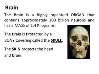
Computed tomography • A cranial computed tomography (CT) scan is an imaging method that uses x-rays to create cross-sectional pictures of the head, including the skull, brain and facial bone.
Contraindication of CT: • Pregnancy except in rare cases such as post trauma. • Contrast allergy Contraindication of contrast media: • Hemorrhage to avoid increasing the area of Hemorrhage.
Head CT scan procedures: • Trauma CT scan. • Routine CT scan: • Plain (c-). • Routine with contrast (c+). • 2D \ 3D.
Trauma CT scan: Indication: • Adult hydrocephalus , headache – stroke. • Acute infarction (CVA). • Hemorrhage trauma. Patient preparation: • Commonly non. • If the patient is not stable , stabilize the patient and sedation as need.
Patient position: • Supine with head first in gantry. Vertical center: on the mid of the head. Horizontal line: on the orbital line. Important planes: • Saggitalline • Optical meatusline • Orbital line.
Technique: • Start with the patient data registration. • lateral scout film (90 degree).
From the scout film, select the start and end location. Start location: gantry tilt line parallel with the canthromeatal around 22 degree. End location: end of the skull use the lateral topogram (scout). • Gantry tilt parallel with optical meatusline, to avoid eyes exposure & to show the anatomy & small lesions within brain fossa (anterior, mid & posterior).
Axial scan: • Thickness of slice x spacing = 5x5 mm continuous scanning
Types of image windowing Reconstruction types: • STANDARD. • BONE.
Routine CT scan: 1- Plain (C-) Indication: • enlarged brain cavities (ventricles) in patients with hydrocephalus. • VP shunt ( drainage of fluids from dilated ventricles in case of hydrocephalus to role out obstruction of VP tube). • Headache. • Brain lesion or tumor. • Epilepsy. • Vertigo. • Dizziness.
Preparation: • NPO 3-4 hrs. • Patient not allergic, not asthmatic. • Renal function test. • Diabetic patients not more than 3 months before the examination. • Non diabetic 6 months. • In patient one week. • Pregnancy test for married female. • Sedation for pediatric patient as needed (≤ 12 years). Procedure: Same as trauma scan.
2- Routine with contrast (C+) Indication: • Metastasis: include the brain and whole body scan. • Brain tumor. Preparation: • Same as before.
Technique: Repeat the same series done for plain, then injects the CM. Types of CM: Omnipaque or xenetix. Amount: • Adult (≥13 YEARS) 50cc IV. • Pediatric weight x 2.
3- 2D &3D Indication: • Microcephalic: small head. • Craniostenosis: enlarged head. • Skull fracture. Technique: • Same as plain but with different technique parameter. • Slice thickness x spacing = 5 x 2.5mm overlap scanning (not missing any lesion).
Therapeutic rule of brain CT Infusion (perfusion): Aim: • earlier diagnosis for infarction (no need to waits 72 hrs after the start of weakness). • Increase the blood supply to the affected area. No. of images: at least 500 image.
Technique: • Brain plain:to localize the area of interest. • Inject the CM with the automatic injector. • Cover 8 cm of interested area. Advantage: Earlier diagnosis gives effective treatment. Disadvantages: High radiation dose ( = 40 chest x-ray).
After care: Ask the patient to drink a lot of fluids. Complication: none. Immobilization devices: Axial holder.
Reference:KKUH THANKYOU..