Download
1 / 44
450 likes | 868 Views
Medical Microbiology. Phagocytosis and the Interactions of Various Phagocytes. BIOL 533 Lecture 6. Leukocyte Chemotaxins. Types of chemotaxins C5a attracts neutrophils and monocytes Made by bacteria

E N D
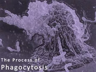
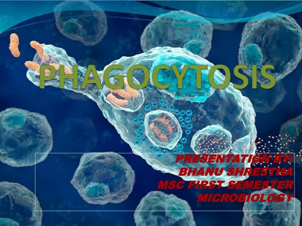
Medical Microbiology Phagocytosis and the Interactionsof Various Phagocytes BIOL 533 Lecture 6
Leukocyte Chemotaxins • Types of chemotaxins • C5a attracts neutrophils and monocytes • Made by bacteria • Peptide clipped off N-terminus (beginning with N-formylmethionine) during peptide maturation after protein synthesis • Made by bacteria and nucleated blood cells • Leucotrienes—lipid products of cell membrane metabolism
Leukocyte Chemotaxins • Function of chemotaxins • Enhance and direct motility of phagocytic cells • To a limited extent, oxidative metabolism of phagocytic cells
Opsonization and Opsonins • General aspects • Substances that enhance ability of phagocytes to ingest microbes • Defend against presence of capsules and other microbial mechanisms that interfere with phagocytosis
Opsonization and Opsonins • Types of opsonins • Antibodies • C3b component of complement • Binds covalently to bacterial surface and is recognized by receptors on neutrophils, monocytes, and macrophage • Bacteria become bound to surface of phagocyte facilitating their uptake
Opsonization and Opsonins • Types of opsonins, continued • Mechanism • White blood cell receptors for C3b • At least 3—CR1, CR2, CR3 (complement receptor) • Children deficient in CR3 very vulnerable to bacterial infections
Phagocytes—Types of Cells • Neutrophils—cell origin • Actively motile cells produced in bone marrow • Differentiate from stem cells over about a two-week period • Production of granules during this time • Azurophil • Produce specific granules later
Phagocytes—Types of Cells • Neutrophils—cell origin, continued • Upon maturation (in numbers of 1010 per day), they move into peripheral blood and circulate for about 6.5 hours • Next move into capillary bed and marginate
Phagocytes—Types of Cells • Neutrophils—cell origin, continued • Margination caused by stickiness due to interleukin-1 • Summoned by chemotaxis, they move through endothelial cell junctions (diapdesis) into extravascular tissue spaces
Phagocytes—Types of Cells • Neutrophils are most active in gut • Gut has enormous microbial population lying just one cell layer away from aseptic tissue • Flora generates large amounts of chemotaxins that recruit most of body’s available leukocytes
Phagocytes—Types of Cells • As a result, submucosa of gut is in a constant state of inflammation • Keep microbial flora down • Synthesis of neutrophils inhibited by chemicals or radiation • Infections in gut region
Phagocytes—Types of Cells • Monocytes and macrophage • Compared to neutrophils • Arrive at damaged tissue later in infection • Days after neutrophils have been active in fighting intruders • Eventually settle in tissues and become resident macrophage
Phagocytes—Types of Cells • Monocytes and macrophage • Share progenitor cell type, but kinetics of maturation & appearance are very different • Monocytes continue cell differentiation after leaving bone marrow • Monocytes and macrophage involved in both constititive and inducible mechanisms • Interact with T cells and play important role in cell-mediated immunity
Phagocytes—Types of Cells • Tissue (resident) macrophage • Exist throughout body • Different names and functions in different tissues • Kupffer cells—liver • Alveolar macrophage—lungs • Osteoclasts—bone • Microglia—brain
Phagocytes—Types of Cells • Monocyte and macrophage functions • Phagocytize invading microbes • Contribute greatly to inflammatory response • Releases • IL-1—enhances sticking of neutrophil to capillary endothelia • TNF—activates newly arrived neutrophils
Mechanism of Phagocyte Killing • Neutrophils • General steps • Attach to microbes • Ingest microbes • Kill microbes • Granules—considered as enlarged lysosomes containing hydrolytic enzymes
Mechanism of Phagocyte Killing • Neutrophil granule types • Azurophil (primary granule) • Contains • Lysozyme • Elastase • A chymotryptic-like protease • Myeloperoxidase • Several antibacterial cationic proteins
Mechanism of Phagocyte Killing • Neutrophil granule types • Specific (secondary granule) • Contains • Cytochrome • Lysozyme • Lactoferin (iron-binding protein) • Vitamin B12 binding protein • Collagenase
Mechanism of Phagocyte Killing • The neutrophil membrane • Contains receptors for chemotaxin and opsonins • After binding chemotaxins, receptors are internalized and replaced with new ones
Mechanism of Phagocyte Killing • Effectiveness of chemotaxis: very effective • Neutrophils are very motile • Move by rearranging cytoplasmic microfilaments and microtubules • Actin and myosin in microfilaments are affected by protein gelsolin • Portions that face upstream in chemotactic gradient form structure called lamellipodium • Cytoplasm is densely packed with microfilaments • Portions face downstream form knob-like uropod
Mechanism of Phagocyte Killing • Process of phagocytosis • General aspects • Differs from pinocytosis in that particles, not liquids, taken up
Mechanism of Phagocyte Killing • Process of phagocytosis, continued • Receptors on phagocyte surface progressively attach to ligands on bacterial surface • Stimulates mechanisms of killing • Oxidative metabolism leading to production of hydrogen peroxide and compounds lethal to microbes (oxygen-dependent killing) • Discharge of toxic compounds from granules into phagosome (oxygen-independent killing)
Mechanism of Phagocyte Killing • Process of phagocytosis, continued • Form phagosome—pouch-like structure that invaginates, displacing the nucleus and granules toward uropod • Form phagolysosome—membrane of granules and phagosome fuse, releasing toxic substances • Forms separate pinched-off organelle • Bacteria coated with antibacterial proteins
Oxygen-Dependent Killing • Fusion of specific granules with phagosome membrane (derived from plasma membrane) brings together: • NADPH oxidase (oxidizes NADPH; found in neutrophil plasma membrane) • Unique cyt b (granule membrane) • A quinone
Oxygen-Dependent Killing • Reaction • O2 O2— (reduces oxygen to superoxide radical) • 2O2—+ H2O H2O2+ O2 (superoxide dismutase)
Oxygen-Dependent Killing • Patients lacking cytochrome components • Children having chronic granulomatous disease (CGD) • Failure to synthesize superoxide radical and therefore hydrogen peroxide • Due to decreased amount of cytochrome b • Gene for larger subunit is missing (90K, 20K)
Oxygen-Dependent Killing • Children having chronic CGD, cont’d. • Neutrophils can phagocytize normally, but do not efficiently oxidize NADPH and kill via oxidative pathway • Usually don’t survive into adulthood
Oxygen-Dependent Killing • How does oxidative process kill? • Interaction with myeloperoxidase supplied by fusion with azurophil • Combines chloride ions and hydrogen peroxide to form hypochlorous ions (analogous to bleach) • Bacteria lacking catalase produce hydrogen peroxide (pneumococci); basically commit suicide • Pneumococci are not dangerous to CGD patients
Oxygen-Independent Killing • Process • Triggered by binding opsonized bacteria to the plasma membrane of neutrophils • Specific granules fuse first • Deliver several bacteriodical proteins, including lysozyme and lactoferin
Oxygen-Independent Killing • Azurophil granules discharge antimicrobial cationic proteins • Some are amphipathic and resemble other cationic surface proteins such as polymyxin B
Oxygen-Independent Killing • Azurophil granules, continued • Disrupt outer membrane of Gram— and kill by causing leakage of vital components • Each of the proteins has unique antimicrobial spectrum, but tend to affect Gram— more than Gram+ • Proteins may account for survival of some CGD children
Oxygen-Independent Killing • Efficiency • Bacterial killing under highly anaerobic conditions of deep abscesses • Patients lacking genes • Coding for cationic proteins • None found, maybe lethal
Oxygen-Independent Killing • Chediak-Higashi syndrome (genetic disease) • Premature fusion of neutrophil granules while cells in bone marrow • When mature cells phagocytize, granules are already spent, substantially reducing killing power
Comparison of Bacterial Sensitivity • Gram— rods in gut killed by oxygen-independent • Gram+ bacteria on skin and upper respiratory epithelia are resistant to oxygen-independent and killed by oxygen-dependent
Mechanism of Phagocyte Killing • Eosinophils • Much like neutrophils, but indicative of parasitic infection
Killing by Monocytes and Macrophage • General aspects • Tend to take care of what is left after battle with neutrophils • Mechanisms of chemotaxis, phagocytosis, and killing resemble mechanisms of neutrophils • Not studied in same detail
Killing by Monocytes and Macrophage • Differences • Continue to differentiate after leaving bone marrow and are activated • Called “angry macrophage” • Phagocytize more vigorously • Take up more oxygen • Secrete large quantity of hydrolytic enzymes • In general, better prepared to kill
Killing by Monocytes and Macrophage • Activated by • Elicited by substances made in response to presence of bacteria (C3b) or viruses (interferon) • Endotoxin of Gram— • Tetrapeptide derived from immunoglobulins (tuftsin)
Killing by Monocytes and Macrophage • Microbial (bacterial, fungi, protozoa) growth within • Some can grow until activated, then killed • Participation in immune response • Help rid body of not only microbial invaders, but also tumor and foreign cells
Killing by Monocytes and Macrophage • Immune response process • Stimulate development of T lymphocytes • Respond to signals from other lymphocytes that stimulate differentiation and activation of macrophage
Phagocytotic Killing • Macrophages/neutrophils/mast cells stimulated by • TNF • interferon • Produce reactive nitrogen intermediates • Nitric oxide • Nitrite (NO2—) • Nitrate (NO3—)
Phagocytotic Killing • Released from cells or contained within vacuoles • Macrophages produce NO from arginine when stimulated by cytokines • NO can block cellular respiration by complexing iron in electron transport proteins
Macrophage Killing • Herpes simplex • Toxoplasma gondii • Leishmania major • Cryptococcus neoformans • Schistosoma mansoni
Lecture 6 • Questions? • Comments? • Assignments...