Download

1 / 40
400 likes | 918 Views
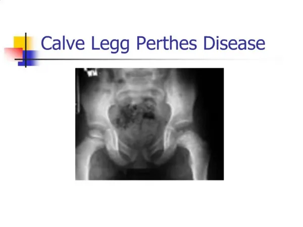
Legg- Calve – Perthes disease. Anatomy. Acetabular retroversion. Etiology of Legg-Calve- Perthes Disease. Factors That May Be Etiologic Trauma Susceptible child Hereditary factors Coagulopathy Hyperactivity Passives smoking Factors Unlikely To Be Etiologic Endocrinopathy

E N D
Etiology of Legg-Calve-Perthes Disease Factors That May Be Etiologic • Trauma • Susceptible child • Hereditary factors • Coagulopathy • Hyperactivity • Passives smoking Factors Unlikely To Be Etiologic • Endocrinopathy • Urban environment • Synovitis
Clinical Features of Legg-Calve-Perthes Disease • Onset: between 18months of age and skeletal maturity (most prevalent between 4-8 years )12years of age • Male sex prevalence: the disease is four or five times more likely to develop in boys than in girls • Involvement: bilateral in 10%to 12% of patients
Clinical Features of Legg-Calve-Perthes Disease Symptoms -limp that is exacerbated by activity and alleviated with rest -pain, which may be located in the groin,anterior hip region, medial knee joint or laterally around the greater Trochanter - history of antecedent trauma
Clinical Features of Legg-Calve-Perthes Disease Signs • Abductor limp • Decreased range of motion of the hip, especially on abduction and internal rotation - Flexion/extension less affected
Symptoms and Signs of Legg-Calve-Perthes Disease Symptoms • Limping • Hip pain • Knee pain • History of trauma (?) Signs • Limp • Decreased hip range of motion • Spasm of long muscle around hip joint
Differential Diagnosis for Legg-Calve-Perthes Disease Other Causes of Avascular Necrosis • Sickle cell disease • Other hemoglobinopathies • Thalassemia • Steroid medication • After traumatic hip fracture & dislocation • Treatment of developmental dysplasia of the hip
Pathologic Findings of Legg-Calve-Perthes Disease Early Stage • Dead trabecular bone , Collapsed trabeculae • Thickened articular cartilage , Physeal disruption • Cartilage extending from the physis into the metaphysis Fragmentation Stage • Invasion of vascular granulation tissue • New bone forming on old trabeculae • Woven new bone formation Healing Stage • New bone, woven and lamellar • Return to normai architecture
Differential Diagnosis for Legg-Calve-Perthes Disease EpiphysealDysplasias • Muitipleepiphyseal dysplasia • Spondyloepiphyseal dysplasia • Mucopolysaccharidoses • Hypothyroidism
Differential Diagnosis for Legg-Calve-Perthes Disease Other Syndromes • Osteochondromatosis • Metachondromatosis • Schwartz-Jam pel syndrome • Trichorhinophalangeal syndrome • Maroteaux-Lamy syndrome
Caterall classification • Group I, partial head or less than half head involvement; • Groups II and III, more than half head involvement and sequestrum formation • Group IV, involvement of the entire epiphysis
Imaging Evaluation • MRI • Bone scan • Arthrography • X-ray
Treatment The primary aim of treatment of Legg-Calv-Perthes disease is containment of the femoral head within the acetabulum
Treatment 1. Most patients can be treated by noncontainment methods and obtain good results (84%).2. Satisfactory clinical results frequently can be obtained at long-term follow-up despite an unsatisfactory radiographic appearance (nine hips).3. The Catterall classification is a valid indicator of results, but is not applicable as a therapeutic guide for an average of 8.1 months after onset
Treatment 4. Head-at-risk signs added little to the Catterall classification as a prognostic indicator or therapeutic guide.5. All of the fair and poor results were in patients with Catterall III or IV involvement and onset of the disease at age 6 or older. (A Catterall III or IV classification is equivalent to Herring groups B and C.)