Download
1 / 44
540 likes | 1.03k Views
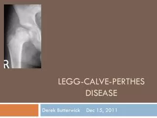
Legg-Calve-Perthes Disease S. Maryam Moghadam. Definition. Idiopathic osteonecrosis of the capital femoral epiphysis of the femoral head of unknown aetiology. It is a self-limited disease. Etiology. Infection, trauma, synovitis Disruption of blood flow to capital femoral epiphysis (CFE)

E N D
Legg-Calve-Perthes Disease S. Maryam Moghadam
Definition Idiopathic osteonecrosis of the capital femoral epiphysis of the femoral head of unknown aetiology. It is a self-limited disease.
Etiology Infection, trauma, synovitis Disruption of blood flow to capital femoral epiphysis (CFE) Systemic disorder (delayed skeletal maturation, abnormalities of thyroid hormone and insulin like growth factor Hereditary influence, environmental influence, hyperactivity
Epidemiology One in 1200 children younger than 15 years is affected by LCPD Males are affected 4-5 times more often than females LCPD most commonly is seen in persons aged 4-8 (2-12) years, with a average age of 7 years Bilateral involvment 10 -15%
Pathology The blood supply to the capital femoral epiphysis is interrupted (arteries and veins). Bone infarction occurs, especially in the subchondral cortical bone, while articular cartilage continues to grow. (Articular cartilage grows because its nutrients come from the synovial fluid.) Revascularization occurs, and new bone ossification starts. Changes to the epiphyseal growth plate occur secondary to the subchondral fracture.
Symptoms Painless limp Hip or groin pain, which may be referred to the thigh Mild or intermittent pain in anterior thigh or knee Usually no history of trauma
Symptoms Decreased range of motion (ROM), particularly with internal rotation and abduction Painful gait Atrophy of thigh muscles secondary to disuse Muscle spasm- mild hip contracture of 10-20 degrees may be present
Symptoms Leg length inequality due to collapse Thigh atrophy: Thigh circumference on the involved side will be smaller than on the unaffected side secondary to disuse (Trendelenburg sign)
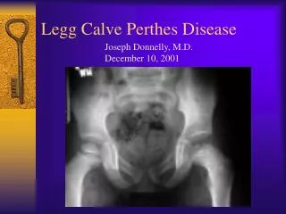
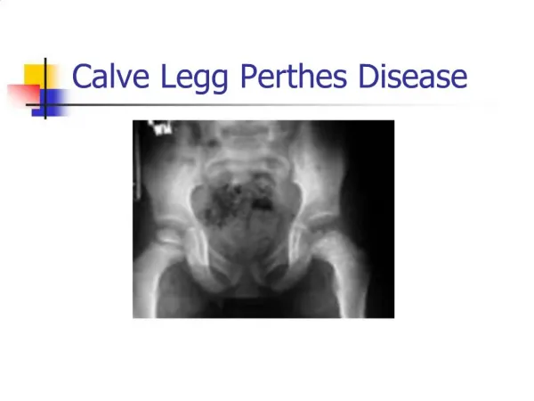
Diagnosis Clinical presentation, physical examination RTG- A-P, frog-leg lateral views (every 6 weeks at the beginning, every 3-6 months later) USG- synovitis MRI, artrography
Stages – radiographic presentation Ischaemia / Necrosis Fragmentation / Resorption Reossification / Healing Residual stage
Initial stage- necrosis Decreased size of ossification center Lateralization of femoral head Subchondral fracture Physeal irregularity
Fragmetation- resorption Fragmented epiphysis More irregular acetabular contour
Reossification- healing New bone formation- the bone density returns
Residual stage Reossified femoral head Remodeling of the head shape Remodeling of the acetabulum
Catterall classification Stage 1: Antero-medial portion of head involved and no collapse, metaphyseal changes do not occur and the epiphyseal plate is not involved Heal without significant sequelae Stage 2: More head involved and may - fragmentation of the involved segment The involved segment shows increased density and uninvolved pillars of normal bone prevent significant collapse - regeneration without much loss of height and the end result is usually good. Metaphyseal reaction localised
Catterall classification Stage 3: More of the head involved - collapse as uninvolved pillars not large enough t prevent collapse May show head within a head The metaphysis is usually diffusely involved - broad neck and the epiphyseal plate is unprotected and also usually involved - results poorer Stage 4: Whole head involvement and severe collapse occurs early and restoration of the femoral head usually less complete The metaphyseal changes may be extensive The epiphyseal plate is often involved - abnormal growth (coxa magna, coxa breva, coxa vara and coxa valga)
Herring classification Lateral pillar classification Determine treatment and prognosis
Salter - Thompson Classification Stage A: - Lateral portion of femoral capital epiphysis present - less than 50% head involved Stage B: - Lateral portion of femoral capital epiphysis absent - more than 50% head involved (Lateral margin of epiphysis protects epiphysis from stress)
Mose method If head conforms to a single ring in both X-Ray planes - good prognosis If head varies from perfect circle by no more than 2mm - fair results If head varies by more than 2mm in any plane - poor results
Centre-edge angle 5-8 years ~19 degrees 9-12 years ~25 degrees 13-20 years 26-30 degrees
Goal of treatment Preservation of the roundness of the femoral head and prevention of deformity while the condition runs its course.
Conservative treatment Relieve weight bearing Achieve and maintain ROM Containment of the femoral epiphysis within the confines of the acetabulum (Petrie-style casts, Atlanta /Scottish Rite/ brace, Toronto braceand other orthotic devices)
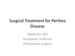
Surgical treatment Femoral osteotomy = varus +/- derotation to reduce the degree of anteversion & extension. Pelvic osteotomy (Salter, Chiari, Shelf) or Femoral osteotomy have similar results
Surgical treatment Shelf acebuloplasty
Surgical treatment Salter osteotomy
Very good radiographic resultsbefore surgery (7 years 2 months)


![[PDF READ ONLINE] The Parents' Guide to Perthes: Understanding Legg-Calvé-Perthes Disease](https://cdn7.slideserve.com/12581636/slide1-dt.jpg)