Download
1 / 63
680 likes | 1.08k Views
Cellular Components of the Immune Response: Stem Cells and Stem Cell Transplantation. Folder Title: Cells Without Turning Point Slides. Updated: October 21, 2013. Filename: CellsNoTP.ppt. Immune System Make-up. From 447Intro, Slides 54 and 55.

E N D
Cellular Componentsof theImmune Response:Stem Cells and Stem Cell Transplantation Folder Title: Cells Without Turning Point Slides Updated: October 21, 2013 Filename: CellsNoTP.ppt
Immune System Make-up From 447Intro, Slides 54 and 55
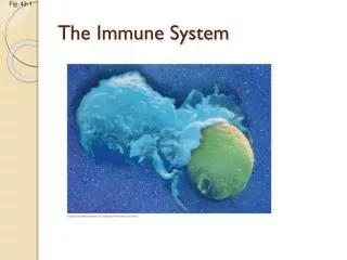
Morphology & Staining of Blood Cells Kuby, 3rd Ed. Figure 3-1 ImmCells
Questions About Cellular Components of the Immune Response How many different cell types are there? What are the numbers of the various cell types? What do these different cell lineages do? Where do they come from? How Long do they last? What becomes of them at the end of their functional life span? What controls their replacement? (How does the hematopoietic system know what needs to be replaced?) What happens if they aren’t replaced correctly? If they are deficient in number? If they are produced in excess to what is needed? If they are not structurally normal?
Blood Counts “RBC” 5x10⁹/ml blood “WBC” 7.3x10⁶/ml blood Ratio RBC to WBC = 685:1 “WBC” = White Blood Cells (Leucocytes) See Table 2-4, p. 30, Kuby 6th Edition
Mouse Whole Blood with Human Leukemia Cells Added at ~0.5% Diluted 1:500 for Counting
Blood Cell Survival Times and Turn-Over Erythrocytes (Red Blood Cells) ~ 4 Months Neutrophils 1 Day Lymphocytes Years White Blood Cell Generation 3.7 x 10¹¹/day(50 x World Human Population per Day)
Blood Cell Replacement Problems At the correct time: When cells are damaged, aged, or no longer functional or necessary. Replace with the correct cell type. In the correct number. Do not propagate errors arising during cell division.
Replacing Cells at the Correct time Getting Rid of Aged or Damaged Cells Without generating inflammation: Genetically Programmed Cells Death (Apoptosis)vs Inflammatory Lysis and Necrosis
Genetically Programmed Cell Death: Apoptosis
Hematopoeisis and Leukemogenesis (Leukemia) What happens if damaged cells are not destroyed? What happens if Apoptosis is not invoked? Bcl-2 gene up-regulation in leucocytes leads to leukemia.(Strong inhibition of Apoptosis) FAS Gene or Caspase Genes down-regulated or lost in cells leads to leukemia and other cancer.(Failure to initiate or promote Apoptosis)
Replacing Cells at the Correct Time with the Correct Cell Type Where Do the Blood Cells Come From? Stem Cells and Partially Differentiated Progenitor Cells Stem Cell Therapy in Medicine
200,000 peripheral blood cells restore viability 1,000 mixed stem cells and progenitor cells restore viability
Pure Stem Cells 30 to 100 Stem cells restore viability See Figure 2-5 Kuby, 6th Ed.
Please put away all notes and any devices except for your Turning Point NXT Transmitter. No papers or computers on your desk please. No communication between or among students.
It takes 200,000 cells to restore viability in the lethally irradiated mouse at the top, but only 1,000 cells are required for the mouse at the bottom? Why is that?
Isolation of CD34+ Pluripotent Stem Cells from Mixed Bone Marrow Mononuclear Cells Using Monoclonal Antibody to CD34 Marker on Stem Cells CD34 Purification of Stem Cells See Figure 3-9 Kuby, 3rd Ed. Antibody conjugated to Biotin. Avidin coats insoluble beads Biotin Sticks Strongly to Avidin
Stem Cell Transplantation in Medicine: In Immune deficiency diseases In immuno-suppressed states Autologous Transplants Syngeneic Donors Allogeneic Donors
Autologous Transplant See p.38 Kuby, 4th Ed.
Pluripotent Stem Cell and Lymphoid and Myeloid Lineages(Fig 2-1, Kuby 4th Ed. p. 28 StemCell Myeloid Stem Cell
Hematopoiesis (formation of blood cells)Fig 2-1, Kuby 4th Ed. p. 28 HematoAll
Lymphoid Lineage Fig 2-1 Kuby 4th Ed p. 28 Lymphoid
Cells of Lymphocyte Lineage B-Lymphocytes: Antibody receptors and antibody production T-Lymphocytes (Thymus-derived lymphocytes): T-Cell Receptors Helper T-Cells “CD4 positive” Cytotoxic T-Cells “CD8 Positive” Natural Killer Cells (“Non-B-Cell, Non-T-Cell Lymphocytes) Recognize virally infected or transformed cells Bind to antibody labelled cells as part of antibody-dependent, cell mediated cytotoxicity (ADCC)
Differentiation Antigen Markers (CD Antigens) on Lymphocytes p. 34 Used to identify sub-populations of lymphocytes and to isolate them
Appendix 1: Pages A1 to A26 339 CD Antigens on Leucocytes What cells types express them What they do e.g. CD4 is a co-receptor on helper T-cells. Confirms binding of T-Cell with its T-Cell Receptor to an antigen-presenting cell.
Natural Killer Cells • Large Granular Lymphoctyes • Part of innate natural immune response • Usually without T-Cell Receptor or Membrane Antibody • Recognize patterns of surface molecules or unusual expression of self-molecules (Class I MHC) • Have anti-tumor and anti-viral activity • CD16 Membrane Receptor for specific antibody regions gives Antibody-dependent cell-mediated cytotoxic activity • NKT Cells have TCR, • Bind to MHC-like molecules CD1 • Secrete cytokines
Hematopoiesis (formation of blood cells)Fig 2-1, Kuby 4th Ed. p. 28 HematoAll
Cells of Myeloid Lineage Polymorphonuclear leukocytes: (Granulocytes) Neutrophils, Eosinophils, Basophils, Mast Cells Antimicrobial, allergic reactions, ADCC Monocyte Macrophages: Mononuclear phagocytes Antimicrobial, attack virally infected cells, Phagocytosis, Endocytosis, & Pinocytosis Degrade and present processed antigens Denedritic Cells Similar functions as for macrophages (Note “Follicular Dendritic Cells, p. 40) Different from bone-marrow derived dendritic cells Erythrocytes: Red Blood Cells, carrying oxygen Megacaryocytes: Produce platelets for blood clotting
Myeloid Lineage Myeloid Lineage (Kuby, Fig 2-1, 4th Ed., p. 28. See Figure 2-2 Hematopoiesis 6th Edition p. 25)
Differentiation in the Monocyte Macrophage Lineage Myeloid to Monocyte
BloodCells2 Histiocyte (Connective Tissue) Mesangial Cell (Kidney) Osteoclast (Bone)
Monocyte to Macrophage Macrophage (MPH or MO) Monocyte Monocyte to Macrophage Kuby, Immunology. 6th Edition Figure 2-7 MC&MPH
Macrophage Ingesting and Degrading Bacterial TargetsKuby, Immunology, 6th Edition, Figure 2-8 Macrophage bacteria Antigen presentation MPHIngest
Kuby, 4th Edition, p. 44 Macrophage Factors MPhMake
Types of Antigen-Presenting Dendritic Cells.
Differentiation and Function in the Granulocyte Lineage Neutrophils: Eosinophils: Basophils and Mast Cells
BloodCells2 Histiocyte (Connective Tissue) Mesangial Cell (Kidney) Osteoclast (Bone)
Differentiation and Function in the Granulocyte Lineage Neutrophils: Multi-lobed Nucleus (PMN) Polymorphonuclear Leucocyte 50% of circulating leukocytes. Short-lived (Hours or Days). Phagocytic Circulates, extravasates out of vasculature into tissue. Responds to chemotactic factors released by infection and inflammation (e.g. from complement or blood-clotting reactions or cytokines released by T-cells or macrophages). Granules release peroxidase, lysozyme, hydrolases, proteases, collagenase. Antimicrobial agents released. Part of innate natural immune response.
Macrophage and PMN Killing Agents Kuby, 4th Edition, p. 43 MPhKill