Download
1 / 9
90 likes | 93 Views
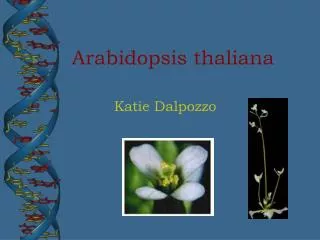
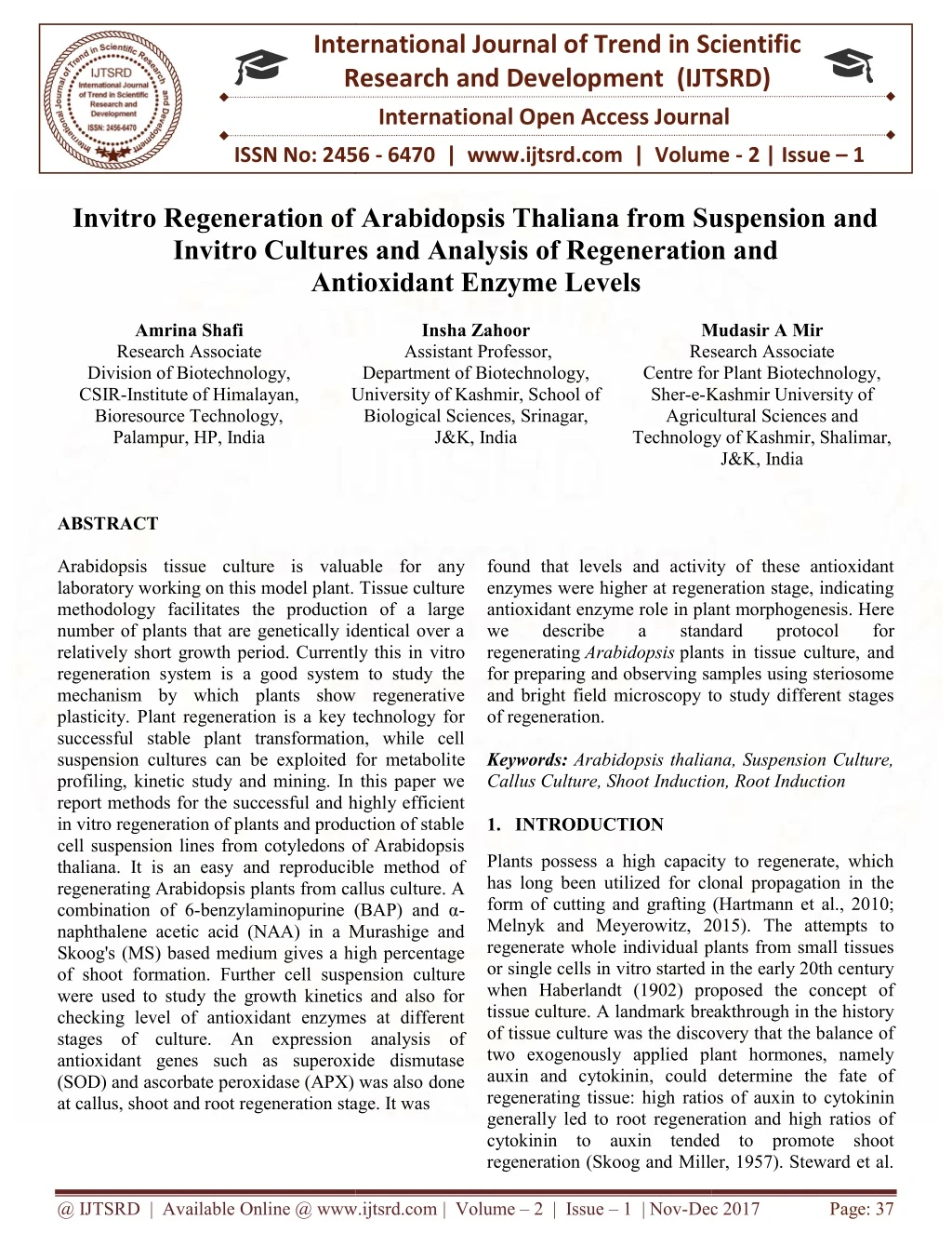
Arabidopsis tissue culture is valuable for any laboratory working on this model plant. Tissue culture methodology facilitates the production of a large number of plants that are genetically identical over a relatively short growth period. Currently this in vitro regeneration system is a good system to study the mechanism by which plants show regenerative plasticity. Plant regeneration is a key technology for successful stable plant transformation, while cell suspension cultures can be exploited for metabolite profiling, kinetic study and mining. In this paper we report methods for the successful and highly efficient in vitro regeneration of plants and production of stable cell suspension lines from cotyledons of Arabidopsis thaliana. It is an easy and reproducible method of regenerating Arabidopsis plants from callus culture. A combination of 6 benzylaminopurine BAP and a naphthalene acetic acid NAA in a Murashige and Skoogs MS based medium gives a high percentage of shoot formation. Further cell suspension culture were used to study the growth kinetics and also for checking level of antioxidant enzymes at different stages of culture. An expression analysis of antioxidant genes such as superoxide dismutase SOD and ascorbate peroxidase APX was also done at callus, shoot and root regeneration stage. It was found that levels and activity of these antioxidant enzymes were higher at regeneration stage, indicating antioxidant enzyme role in plant morphogenesis. Here we describe a standard protocol for regenerating Arabidopsis plants in tissue culture, and for preparing and observing samples using steriosome and bright field microscopy to study different stages of regeneration. Amrina Shafi | Insha Zahoor | Mudasir A Mir "Invitro Regeneration of Arabidopsis Thaliana from Suspension and Invitro Cultures and Analysis of Regeneration and Antioxidant Enzyme Levels" Published in International Journal of Trend in Scientific Research and Development (ijtsrd), ISSN: 2456-6470, Volume-2 | Issue-1 , December 2017, URL: https://www.ijtsrd.com/papers/ijtsrd5843.pdf Paper URL: http://www.ijtsrd.com/biological-science/biotechnology/5843/invitro-regeneration-of-arabidopsis-thaliana-from-suspension-and-invitro-cultures-and-analysis-of-regeneration-and--antioxidant-enzyme-levels/amrina-shafi<br>

E N D
International Research Research and Development (IJTSRD) International Open Access Journal ro Regeneration of Arabidopsis Thaliana from Suspension and Invitro Cultures and Analysis of Regeneration and Antioxidant Enzyme Levels International Journal of Trend in Scientific Scientific (IJTSRD) International Open Access Journal ISSN No: 2456 ISSN No: 2456 - 6470 | www.ijtsrd.com | Volume www.ijtsrd.com | Volume - 2 | Issue – 1 Invitro Regeneration of Arabidopsis Thaliana from Suspension and Invitro Cultures and Analysis of R Antioxidant Enzyme L Amrina Shafi Research Associate Division of Biotechnology, CSIR-Institute of Himalayan, Bioresource Technology, Palampur, HP, India ro Regeneration of Arabidopsis Thaliana from Suspension and egeneration and Insha Zahoor Assistant Professor, Department of Biotechnology, Mudasir A Research Associate Centre for Plant Biotechnology, Sher-e-Kashmir University of Agricultural Sciences and Technology of Kashmir, Shalimar, Technology of Kashmir, Shalimar, J&K, India Mudasir A Mir Research Associate Centre for Plant Biotechnology, Kashmir University of Agricultural Sciences and University of Kashmir, School of University of Kashmir, School of Biological Sciences, Srinagar, J&K, India found that levels and activity of these antioxidant enzymes were higher at regeneration stage, indicating antioxidant enzyme role in plant morphogenesis. we describe a standard regenerating Arabidopsis plants in tissue culture, and for preparing and observing samples using steriosome and bright field microscopy to study different stages of regeneration. Keywords: Arabidopsis thaliana, Suspension Culture, Callus Culture, Shoot Induction, Root Induction 1.INTRODUCTION ABSTRACT Arabidopsis tissue culture is valuable for any laboratory working on this model plant. Tissue culture methodology facilitates the production of a large number of plants that are genetically identical over a relatively short growth period. Currently this in regeneration system is a good system to study the mechanism by which plants show regenerative plasticity. Plant regeneration is a key technology for successful stable plant transformation, while cell suspension cultures can be exploited for metaboli profiling, kinetic study and mining. In this paper we report methods for the successful and highly efficient in vitro regeneration of plants and production of stable cell suspension lines from cotyledons of Arabidopsis thaliana. It is an easy and reproducible method of regenerating Arabidopsis plants from callus culture. A combination of 6-benzylaminopurine (BAP) and α naphthalene acetic acid (NAA) in a Murashige and Skoog's (MS) based medium gives a high percentage of shoot formation. Further cell suspe were used to study the growth kinetics and also for checking level of antioxidant enzymes at different stages of culture. An expression analysis of antioxidant genes such as superoxide dismutase (SOD) and ascorbate peroxidase (APX) was also at callus, shoot and root regeneration stage. It was Arabidopsis tissue culture is valuable for any laboratory working on this model plant. Tissue culture methodology facilitates the production of a large number of plants that are genetically identical over a relatively short growth period. Currently this in vitro regeneration system is a good system to study the mechanism by which plants show regenerative plasticity. Plant regeneration is a key technology for successful stable plant transformation, while cell suspension cultures can be exploited for metabolite profiling, kinetic study and mining. In this paper we report methods for the successful and highly efficient in vitro regeneration of plants and production of stable cell suspension lines from cotyledons of Arabidopsis found that levels and activity of these antioxidant enzymes were higher at regeneration stage, indicating antioxidant enzyme role in plant morphogenesis. Here we describe a standard plants in tissue culture, and for preparing and observing samples using steriosome and bright field microscopy to study different stages protocol protocol f for Arabidopsis thaliana, Suspension Culture, Induction, Root Induction Plants possess a high capacity to regenerate, which has long been utilized for clonal propagation in the form of cutting and grafting (Hartmann et al., 2010; Melnyk and Meyerowitz, 2015). The attempts to regenerate whole individual plants from small tissues or single cells in vitro started in the early 20th century when Haberlandt (1902) proposed the concept of tissue culture. A landmark breakthrough in the history of tissue culture was the discovery that the balance of two exogenously applied plant hormones, namely auxin and cytokinin, could determine the fate of regenerating tissue: high ratios of auxin to cytokinin generally led to root regeneration and high ratios of cytokinin to auxin tended to promote shoot regeneration (Skoog and Miller, 1957). Steward et al. regeneration (Skoog and Miller, 1957). Steward et al. Plants possess a high capacity to regenerate, which has long been utilized for clonal propagation in the form of cutting and grafting (Hartmann et al., 2010; , 2015). The attempts to regenerate whole individual plants from small tissues or single cells in vitro started in the early 20th century when Haberlandt (1902) proposed the concept of tissue culture. A landmark breakthrough in the history e was the discovery that the balance of two exogenously applied plant hormones, namely auxin and cytokinin, could determine the fate of regenerating tissue: high ratios of auxin to cytokinin generally led to root regeneration and high ratios of o auxin tended to promote shoot ucible method of regenerating Arabidopsis plants from callus culture. A benzylaminopurine (BAP) and α- naphthalene acetic acid (NAA) in a Murashige and Skoog's (MS) based medium gives a high percentage of shoot formation. Further cell suspension culture were used to study the growth kinetics and also for checking level of antioxidant enzymes at different stages of culture. An expression analysis of antioxidant genes such as superoxide dismutase (SOD) and ascorbate peroxidase (APX) was also done at callus, shoot and root regeneration stage. It was @ IJTSRD | Available Online @ www.ijtsrd.com @ IJTSRD | Available Online @ www.ijtsrd.com | Volume – 2 | Issue – 1 | Nov-Dec Dec 2017 Page: 37
International Journal of Trend in Scientific Research and Development (IJTSRD) ISSN: 2456-6470 (1958) further demonstrated that even single cells from carrot vascular phloem retain totipotency – the capacity to regenerate whole plants – thus highlighting the astonishing regenerative potential of plant somatic cells. 2010).Furthermore, it has been reported that the cytosolic Cu/Zn- SOD was induced in regenerating tobacco protoplasts (Papadakis et al. 2001), which supports the hypothesis that SOD is involved in plant morphogenesis. The present study was aimed to study callus growth and in vitro regenerative capacity of the different tissues of Arabidopsis. We also showed expression and activity of antioxidant genes has a role to play in plant morphogenesis. Our results suggest that proper maintenance of redox homeostasis is crucial for successful regeneration at the early stages of shoot organogenesis. II. MATERIALS AND METHODS Plant growth, callus induction, and regeneration Arabidopsis seeds were surface sterilized, sown in petri dishes containing Murashige and Skoog (MS)medium. The plates were kept at 4 °C for 2 days and then shifted to 21±1 °C. After 10 days of germination, cotyledons were excised from seedlings and cut into two pieces and placed on MS callus induction medium containing 30 g l−1 sucrose and 8.0 g l−1 agar (Sigma-Aldrich, St. Louis, MO, USA), supplemented with dichlorophenoxyacetic acid (2,4-D) (Sigma-Aldrich, St. Louis, MO, USA). The explants were cultured in the dark at 25±2 °C. Callus formation started after 1 week of inoculation on callus induction medium. After 1 month, compact proliferating callus was selected and transferred to fresh MS medium with 1.0 mg l−1 2,4-D, 30 g l−1 sucrose, and 8.0 g l−1 agar. After 1-month subculture, proliferating callus was transferred to regeneration medium (half strength MS medium with 30 g l−1 sucrose, 8.0 g l−1 agar, supplemented with 0.5–1.0 mg l−1 2,4-D, and 0.5–1.0 mg l−1 benzylaminopurine (BAP) (Sigma-Aldrich, St. Louis, MO, USA) for regeneration in a culture room with a 16-h photoperiod (60–70 μmol m−2 s−1 cool white fluorescent irradiance) for 4 weeks at 25±2 °C. Callus with clearly differentiated shoots were counted as regenerated, with each piece of callus counted as one unit. After 4 weeks, the regenerated plantlets were transferred to 100-ml flasks containing the same medium for further growth. SOD activity and H2O2 content were determined at different stages of culture. Regeneration frequency was calculated as the number of regenerated explants per total number of cultured explants. Three replications were used in each experiment. A common mode of plant regeneration both in nature and in vitro is de novo organogenesis, in which plant cuttings or explants first form ectopic apical meristems and subsequently develop shoots and roots. Meristems are specialized plant tissues where new cells, tissues and organs are generated through cell division and differentiation. Plants can also regenerate through somatic embryogenesis in vitro, whereby isolated protoplasts or cells first develop cellular structures similar to subsequently generate whole plant bodies. Both of these regeneration processes occur either directly from parental tissues or indirectly via the formation of a callus. Over recent decades, various culture conditions have been regeneration and utilized for clonal propagation and genetic transformation in diverse plant species. The regenerative capacity of plant cells can be enhanced in vitro when explants are cultured on nutrient media supplemented with plant hormones (Skoog and Miller, 1957; Murashige, 1974; George et al., 2008)root regeneration can also be induced de novo from various mature somatic tissues, and whole plants can be regenerated even from single protoplasts through de novo organogenesis embryogenesis (Takebe et al., 1971; Zhu et al., 1997; Chupeau et al., 2013). Regeneration process is controlled by various genes whose expression is governed by certain physical and chemical conditions (Imani et al. 2001; Papadakis et al. 2001). So far, in vitro regeneration has been achieved by a variety of means including treatment with plant growth regulators (PGR), temperature shocks, osmotic stress, and through application of various chemical substances (Szechynska-Hebda et al. 2007; Touraev et al. 1997; Zavattieri et al. 2010). The consequence of these processes has been generally considered as ROS overproduction, which is detrimental for plant as such (Scandalios 1997). However, recent pieces of evidence suggest that ROS participate in signal transduction cascade (Prasad et al. 1994) and have a positive role in plant growth and development (Tian et al. 2003). A change in the activity of antioxidant enzymes has also been detected during in vitro shoot initiation and development (Batkova zygotic embryos and established for plant 1 mg l−1 2,4- or somatic et al. 2008; Gupta @ IJTSRD | Available Online @ www.ijtsrd.com | Volume – 2 | Issue – 1 | Nov-Dec 2017 Page: 38
International Journal of Trend in Scientific Research and Development (IJTSRD) ISSN: 2456-6470 estimated according to the dye binding method of Bradford using BSA as standard (Bradford 1976). RT-PCR Analysis Total RNAwas isolated from transgenic and the wild- type Arabidopsis plants using Total RNA Extraction Kit (RealGenomics). One microgram of total RNA was used for oligo (dT) primed first-strand cDNA synthesis in 20-μl reactionusing Superscript III reverse transcriptase (Invitrogen). This cDNA was used in 27-cycle PCR using gene specific primers for PaSOD gene. Constitutively expressed 26S rRNA gene was amplified simultaneously in 27 cycles to ensure equal amounts of template cDNA used. Statistical analysis All experiments were conducted with at least threeindependent repetitions in triplicate. All values areshown as the mean±standard deviation. The statisticalanalysis was performed using Statistica software (v.7).The statistical significance between the mean values was assessed by Analysis of Variance (ANOVA) applying Duncan’s multiple range test (DMRT). A probability level of P≤0.05 was considered significant. III. RESULTS AND DISCUSSION Callus Induction, Initiation of Suspension culture and Growth Kinetic Study In the present study, cotyledons of Arabidopsis plants were used as explants due to their high regeneration potential as suggested by various previous studies (Ozcan et al. 1992; Patton and Meinke 1988; Mante et al. 1989). In addition to this root and stem section of Arabidopsis was also used to induce callus. After 1 week of inoculation on 1 mg l−12,4 D, callus formation has initiated. Callus Induction was faster in cotyledons (CT) followed by root (RT) and stem sections (IF). Callus initiation is the primary stage in many tissue culture processes for the establishment of cell suspension cultures(Kumar and Kanwar 2007; Ngara et al. 2008). Once callus was initiated under in- vitro conditions, inoculum was used for suspension culture (Liquid MS, supplemented with 1 mg l−12,4 D). The growth of callus was determined based on the fresh and dry weights after 10, 20 and 20 days of suspension culture.Growth kinetics showed a typical curve with lag phase, exponentialphase followed by stationary phase after incubation.The pattern of the growth curve obtained in CT, RT and IF was different (Fig.1). Upon transferring the calli ofCT and (RT and Measurement of callus growth Following callus induction after 3 weeks of culture, callus was aseptically transferred onto liquid medium in culture flasks. The flasks were incubated in the dark at 27±1 °C for 4 weeks. Growth kinetics was studied by determination of fresh and dry weights of fresh callus of 5, 10, 15, 20, 25, and 30 days old. Dry weight of fresh callus was determined after drying in a vacuum oven at 65 °C until constant weight. The cultures were incubated for 16-h photoperiods at 25 °C. Microscopy Explants were collected at 0, 1, 2, 3, and 4 weeks from initiation and evaluated using scanning electron microscopy (SEM) and light microscopy (LM). Samples were fixed informalin, glacial acetic acid, and 50 % ethyl alcohol (FAA)(1:1:18) at room temperature. Samples were subsequently dehydrated in a tertiary butyl alcohol series, embedded in paraffin (melting point 58–60 °C), and 8–10-mm thick sections were cut using a Finesee microtome. Sections were stained with 1 % safranin in water and with 4 % fast green in clove oil for 4 h and for 30 s, respectively. These were mounted in Canada balsam and examined using bright fieldmicroscope (Zeiss LSM510 meta GmbH, Germany) equipped with a Zeiss Axiovert 100 M inverted microscope. SOD and APX enzyme activity assay Leaf samples (100 mg) were homogenized in a pre cooledmortar in homogenizing buffer containing 2 mM EDTA,1 mM DTT, 1 mM PMSF, 0.5 % (v/v) Triton-X100 and10 % (w/v) PVPP in 50 mM phosphate buffer pH homogenizing buffer contained ascorbatein addition and the buffer pH was set to 7.0. Thehomogenate was transferred to 1.5 ml tubes and centrifugedat 13,000 rpm for 20 min at 4 °C. The supernatant was collected and total SOD and APX activities were estimated. The total SOD activity was measured by adding5 μl enzyme extract to reaction mixture (200 μl) containing1.5 μm riboflavin, 50 μm NBT, 10 mM Dl- methionine and 0.025 % (v/v) triton-X100 in 50 mM phosphate buffer. One unit of enzyme activity was defined as the amount of enzyme required for 50 % inhibition of NBT reduction per min at 25 °C. Specific activity of SOD was calculated accordingly. APX activity was determined by following the oxidation rate of ascorbate at 290 nm as described by Nakano and Asada (1981). Protein content was 7.8.For APX activity, @ IJTSRD | Available Online @ www.ijtsrd.com | Volume – 2 | Issue – 1 | Nov-Dec 2017 Page: 39
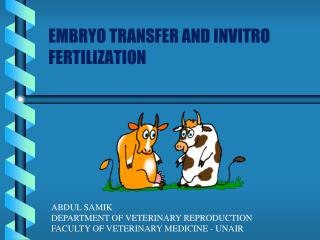
International Journal of Trend in Scientific Research and Development (IJTSRD) ISSN: 2456-6470 IF) to the suspension medium, verylittle increase in biomass was observed during the first 4 daysof culture (the lag phase). After 6 days of culture, the calliwere found in their exponential phase as the cells rapidlydivided and proliferated. After 14 days, culture reached thestationary phase. exponential phase began after 14 days of incubation and stationary phase was reached after22 days. Growth rates during the exponential phase in IF, RTand CTwere 0.22, 0.57, and 0.73 g (dry weight)/day, respectively, (Fig.1 ) while on the basis of fresh weight,growth rates during the exponential phase in IF, RT and CTwere 0.38, 0.98, and 1.22 g (fresh weight)/day, respectively(Fig. 1). These results clearly demonstrate that growthrate of CT calli was significantly higher than that of RT and IFcalli. While in WT, Fig.1 Growth curve analysis of callus from different tissues cotyledons (CT) , root (RT) and Inflorescence (IF) based on d dry weight (DW mg/l) and e fresh weight (FW mg/l), which were determined every 2 days after inoculation. Bar indicates the standard deviation (n=3). Invitro Regeneration Frequency on different concentration of BAP and NAA Calli from IF, RT and CT regenerated shoots when cultured on the regeneration media with different concentrations of BAP and NAA (Table 1). Transfer of callus to regeneration media led to the formation of meristemoids. The meristemoids (nodules or growth centers) are localized clusters of cambium-like cells which may become vascularized due to the appearance of tracheid cells in the center. Formation of meristemoids in callus cultures may represent their association with an early stage development of shoot bud (Kulchetscki et al. 1995). However, an early shoot initiation response was observed from the CT as compared to IF and RT calli on the regeneration medium containing 0.5 mg l−1 NAA and 1.0 mg l−1 BAP. After 2 weeks on the regeneration medium, shoot meristem appeared in approximately 70 % CTcalli (Table 1). Table 1: Callus induction and shoot regeneration from Arabidopsis Cotyledons (CS), Roots (RT) and Inflorescence (IF) tissues Tissue s MS1 MS2 MS3 MS4 RT 5.36±0.06 h CS 7.92±0.09c 13.8±0.13a 5.94±0.06 e d IF 5.94±0.06f 11.1±0.13 b Average diameter (mm) of calli in MS media Percent shoot regeneration in primary calli MS1 25.1±0.48h MS2 30.92±0.48f19.92±0.48i15.18±0.48j MS3 MS4 7.1±0.11d 4.36±0.09i4.06±0.06i 7.08±0.13 60.16±0.65 c 80.08±0.85a 35.08±0.48 36.92±0.48 d 28.16±0.32 g e 29.6±0.31f 5.4±0.06g 5.82±0.08f 34.9±0.70e 62.87±1.22 b @ IJTSRD | Available Online @ www.ijtsrd.com | Volume – 2 | Issue – 1 | Nov-Dec 2017 Page: 40
International Journal of Trend in Scientific Research and Development (IJTSRD) ISSN: 2456-6470 Each value represents mean ± SE of five replicates for each parameter. The induction media contained 1 mg l−1 2, 4-D and regeneration media supplemented with BAP and NAA in different concentrations, MS1 (0.5 mg l−1 NAA/0.5 mg l−1 BAP), MS2 (0.5 mg l−1 NAA/1 mg l−1 BAP), MS3 (1 mg l−1 NAA/0.5 mg l−1 BAP) and MS4 (1 mg l−1 NAA/1 mg l−1 BAP). In each section of the table, means were compared with ANOVA and data followed by the same letters within the columns are not significantly different at the level of P≤0.05, as determined by a least-significant difference (LSD) Expression analysis of antioxidant genes during different Stages of Culture RT-PCR expression analysis of SOD and APX enzymes at callus stage (CS), shooting stage (SS) and rooting Stage (RS) was done (Table 2). It was observed that expression of these gene were induced at SS and RS stages, indicating role of SOD and APX genes in shoot and root regeneration (Fig. 2).Numerous studies have been reported relating to the variation in the patterns of the antioxidant enzyme activity during different stages of organogenesis(Franck et al. 1998; Chen and Ziv 2001; Racchi et al. 2001;Meratan et al. 2009; Vatankhah et al. 2010). Table 2: Primer sequence, PCR conditions, and amplicon size for the PaSOD and 26S rRNA (reference gene) used for semi quantitative PCR Genes Sequence 5’ to 3’ PCR Conditions Amplicon Size (bp) 456 F: TGCCATGGCGAAAGGAGTTGCAG R:ATAGATCTGCGCCCTGGAGACCAATGATG 94 °C, 4 min; 94 °C, 1 min, 56 °C, 30 s, 72 °C, 1 min, 27 cycles; 72 °C, 7 min 94 °C, 4 min; 94 °C, 1 min, 57 °C, 30 s, 72 °C, 1 min, 27 cycles; 72 °C, 7 min 94 °C, 4 min; 94 °C, 1 min, 57 °C, 30 s, 72 °C, 1 min, 27 cycles; 72 °C, 7 min AtSOD F: ATAGATCTGATGGCTGCACCGATTGTT R: TAAGTAGTCTTCATCCTCTTCCGGATCTC 861 AtAPX 26S rRNA F:CACAATGATAGGAAGAGCCGAC R:CAAGGGAACGGGCTTGGCAGAATC 534 Fig.2 Expression analysis of callus stage (CS), Shooting stage (SS) and Rooting Stage (RS). 26S rRNA was used as the loading control. Enzyme Activity Assay of SOD and APXduring different Stages of Culture In the present study, SOD and APX activity was found to vary among SS (shoot stage), RS (root stage) and CS (callus stage) tissues at each stage of culturing. SOD and APX levels were found to be decreased in CS comparing to that of SS and RS stage, then increased in regenerated shoots and roots (Fig. 3). In the case of RS and CS, SOD and APX activity was found to be higher than that of CS at all the stages. However, the increase in the SOD and APX activity of RS and CSlines as compared to WT was more during the regeneration stage. Similar @ IJTSRD | Available Online @ www.ijtsrd.com | Volume – 2 | Issue – 1 | Nov-Dec 2017 Page: 41
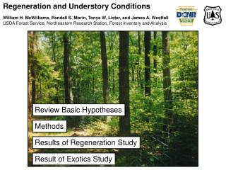
International Journal of Trend in Scientific Research and Development (IJTSRD) ISSN: 2456-6470 observations have also been reported earlier by few groups (Bagnoli et al. 1998; Cui et al. 1999). Microscopic Study of different Stages of Invitro regeneration Under sterosome, different stages of invitro regeneration of Arabidopsis observed. After 1 week of inoculation on 2,4D, callus formation was observed (Fig. 5). Callus turned into green meristemoid like structures after 3 weeks of inoculation of callus on MS media supplemented with BAP and NAA. Theses meristemoid like structures started regenerating leaf primordia like structures after 4 to 4.5 week period of inoculation. After completion of 5 week shoo regeration were observed from the cotyledonary explants (Fig. 5). cotyledons were Fig.5 Micrographs observed under sterosome of Callus induction, Shoot meristemoids formation and shoot Induction in media supplemented by 2,4 D followed by BAP and NAA. Acknowledgments REFERENCES [1]Alscher RG, Erturk N, Heath LS (2002) Role of superoxide dismutases (SODs) in controlling oxidative stress in plants. J Exp Bot 53:1331– 1341 This work was supported by grants from the Councilof Scientific and Industrial Research (CSIR), New Delhi, India, in theformof Network Projects PlaGen (BSC0107) and SIMPLE (BSC0109) atthe CSIR-IHBT. As acknowledge fellowships awarded by the CSIR, India. [2]Arnon D (1949) Copper enzymes in isolated chloroplasts. polyphenol oxidase in Beta vulgaris. Plant Physiol 24:1–14 @ IJTSRD | Available Online @ www.ijtsrd.com | Volume – 2 | Issue – 1 | Nov-Dec 2017 Page: 42
International Journal of Trend in Scientific Research and Development (IJTSRD) ISSN: 2456-6470 [3]Asada K (1994) Production and action of active oxygen species in photosynthetic tissues, causes of photooxidative stress and amelioration of defense systems in plants, In: Foyer CH, Mullineaux PM (eds), CRC Press, Boca Raton, FL, pp 77–104 stress tolerance and improve shoot regeneration. Electron J Biotech 12:1 – 8. [14]Chen J, Ziv M (2001) The effect of an cymidol on hyperhydricity, regeneration, starch and antioxidant enzymatic activities in liquid- cultured Narcissus. Plant Cell Rep 20:22–27 [4]Badawi GH, Yamauchi Y, Shimada E, Sasaki R, Kawano N, Tanaka K, Tanaka K (2004) Enhanced tolerance to salt stress and water deficit by overexpressing superoxide dismutase in tobacco (Nicotiana tabacum) chloroplasts. Plant Sci 166:919–928 [15]Cui K, Xing G, Liu X, Xing G, Wang Y (1999) Effect of hydrogen peroxide on somatic embryogenesis of Lycium barbarum L. Plant Sci 146:9–16 [16]Foyer CH, Descourvierse P, Kunert KJ (1994) Protection against oxygen radicals: an important defense mechanism studied in transgenic plants. Plant Cell Environ 17:507–523 [5]Bagnoli F, Capuana M, Racchi ML (1998) Developmental changes superoxide dismutase in zygotic and somatic embryos of horse chestnut. Aust J Plant Physiol 25:909–913 of catalase and [17]Foyer CH, Lopez-Delgado H, Dat JF, Scott IM (1997) Hydrogen peroxide and glutathione- associated mechanisms of acclimatory stress tolerance and signaling. Plant Physiol 100:241– 254 [6]Barrs HD, Weatherley PE (1962) A re- examination of the relative turgidity technique for estimating water deficits in leaves. Aust J Biol Sci 15:413–428 [18]Franck T, Kevers C, Penel C, Greppin H, Housman H, Gaspar T (1998) Reducing properties and markers of lipid peroxidation in normal and hyperhydrating shoots of Prunu savium L. J Plant Physiol 153:339–346 [7]Bassuner BM, Lam R, Lukowitz W, Yeung E C (2007) Auxin and root initiation in somatic embryos of Arabidopsis. Plant Cell Rep 26:1–11 [8]Bates L, Waldren R, Teare ID (1973) Rapid determination of free proline for water-stress studies. Plant Soil 39:205–207 [19]Gill T, Sreenivasulu Y, Kumar S, Ahuja PS (2010) Over-expression of superoxide dismutase exhibits lignification of vascular structures in Arabidopsis thaliana. J Plant Physiol 167:757– 760 [9]Batkova P, Pospisilova J, Synkova H (2008) Production of reactive oxygen species and development of antioxidative systems during in vitro growth and ex vitro transfer. Biol Plant 52:413–422 [20]Gupta D (2010) Role of free radicals and antioxidants in in vitro morphogenesis. In: Gupta D (ed) Reactive oxygen species and antioxidants in higher plants. CRC Press, pp 230–247 [10]Bechtold N, Ellis J, Pelletier G (1993) In planta Agrobacterium mediated gene transfer by infiltration of adult Arabidopsis thaliana plants, CR Academy of Sciences Paris, Life Sciences 316:1194–99 [21]Halder KP, Burrage SW (2003) Drought stress effects on water relations of rice grown in nutrient film technique. Pakistan J Biol Sci 6: 441–446 [11]Blokhina O, Violainen E, Fagerstedt KV (2003) Antioxidants, oxidative damage and oxygen deprivation stress. a review. Ann Bot 91:179– 194 [22]Hasegawa PM, Bressan RA, Zhu JK, Bohnert HJ (2000) Plant cellular and molecular responses to high salinity. Annu Rev Plant Biol 51: 463–499. [23]Imani J, Tran Thi L, LangenG, Arnholdt-Schmitt B, Roy S, Lein C, Kumar A, Neumann KH (2001) Somatic embryogenesis and DNA organization of genomes from selected Daucus species. Plant Cell Rep 20:537–541 [12]Bohnert HJ, Jensen RG (1996) Strategies for engineering water stress tolerance in plants. Trends Biotechnol 14: 89–97. [13]Chatzidimitriadou Madesis P, Perl-Treves R, Tsaftaris A(2009) Expression of SOD transgene in pepper confer K, Nianiou-Obeidat I, @ IJTSRD | Available Online @ www.ijtsrd.com | Volume – 2 | Issue – 1 | Nov-Dec 2017 Page: 43
International Journal of Trend in Scientific Research and Development (IJTSRD) ISSN: 2456-6470 [24]Klaus A, Heribert H (2004) Reactive oxygen species: metabolism, oxidative stress, and signal transduction. Annu Rev Plant Biol 55:373–399 sativa L.) cultivars differing in salinity resistance. Ann Bot 78:389–398 [35]Madden JI, Jones CS, Auer CA (2005) Modes of regeneration in (Geraniaceae) and three closely related species. In Vitro Cell Dev Biol Plant 41:37–46 [25]Koster accumulation and compartmentation during the cold acclimation of puma rye. Plant Physiol 98:108–113 KK, Lynch DV (1992) Solute Pelargonium hortorum [36]McKown R, Kuroki G, Warren G (1996) Cold responses of Arabidopsis mutants impaired in freezing tolerance. J Exp Bot 47:1919–1925 [26]Kulchetscki L, Harry IS, Yeung EC, Thorpe TA (1995) In vitro regeneration of Pacific silver fir (Abies amabilis) analysis of shoot formation. Tree Physiol. 15:727–738. plantlets andhistological [37]Munns R, Tester M (2008) Mechanisms of salinity tolerance. Annu Rev Plant Biol 59: 651– 681 [27]Kumar N, Bhatt RP (2006). Transgenics: An emerging approach for cold tolerance to enhance vegetables production in high altitude areas. Indian J Crop Sci 1: 8-12 [38]Mante S, Scorza R, Cordts J (1989) A simple, rapid protocol for development from mature cotyledons of Glycine max cv Bragg. In Vitro Cell Dev Biol 25: 385– 388 adventitious shoot [28]Kumar S, Kanwar JK (2007) Plant regeneration from cell suspension in Gerbera gamesonii Bolus. J Fruit Ornam Plant Res 15: 157–166 [39]Matysik J, Bhalu AB, Mohanty P (2002) Molecular mechanisms ofquenching of reactive oxygen species by proline under stress in plant. Curr Sci 82:525–532 [29]Kumar S, Sahoo R, Ahuja PS (2002) Isozyme of autoclavable superoxide dismutase (SOD), a process for the identification and extraction of the SOD in cosmetic, food and pharmaceutical compositions. US Patent No. 6,485950 B1. [40]Meratan AA, Ghaffari SM, Niknam V (2009) In vitro organogenesis and antioxidative enzymes activity in Acanthophyllum sordidum. Biol Plant 53:5–10 [30]Kumar V, Shriram V, KaviKishor PB, Jawali N, Shitole MG (2010) accumulation and salt stress tolerance of transgenic indica rice by over-expressing P5CSF129A gene. Plant Biotechnol Rep 4:37– 48 Enhanced proline [41]Murashige T, Skoog F (1962) A revised medium for rapid growth and bio assays with tobacco tissue cultures. Physiol Plant 15:473–497 [42]Ngara R, Rees J, Ndimba BK (2008) Establishment of sorghum cell suspension culture system for proteomics studies. Afr J Biotechnol 7:744–749 [31]Libik M, Konieczny R, Pater B, Slésak I, Miszalski Z (2005) Differences in the activities of some antioxidant enzymes and in H2O2 content during rhizogenesis embryogenesis in callus cultures of the ice plant. Plant Cell Rep 23:834–841 [43]Ozcan S, Barghchi M, Draper J (1992) High- frequency adventitious shoot regeneration from immature cotyledons of pea (Pisum sativum L). Plant Cell Rep 11:44–47 and somatic [32]Lopez AD, Ahmad OB, Guillot M, Ferguson BD, Salomon JA, Murray CJL, Hill KH (2002) World mortality in 2000: Life tables for 191 countries. Geneva WHO [44]Papadakis (2002) Oxidative stress could be responsible for the recalcitrance of plant protoplasts. Plant Physiol Biochem 40:549–559 AK, Roubelakis-Angelakis KA [33]Luo JP, Jiang ST, Pan LJ (2001) Enhanced somatic embryogenesis by salicylic acid of Astragalus adsurgens Pall: relationship with H2O2 production and H2O2-metabolizing enzyme activities. Plant Sci 161:125–132 [45]Papadakis Angelakis KA (2001) Reduced activity of antioxidant machinery suppression of totipotency in plant protoplasts. Plant Physiol 126:434–444 AK, Siminis CI, Roubelakis- is correlated with [34]Lutts S, Kinet JM, Bouharmont J (1996) NaCl- induced senescence in leaves of rice (Oryza @ IJTSRD | Available Online @ www.ijtsrd.com | Volume – 2 | Issue – 1 | Nov-Dec 2017 Page: 44
International Journal of Trend in Scientific Research and Development (IJTSRD) ISSN: 2456-6470 [46]Patton D, Meinke D (1988) High-frequency plant regeneration from cultured cotyledons of Arabidopsis thaliana. Plant Cell Rep 7:233–237. [56]Shi H, Lee BH, Wu SJ, Zhu JK (2003) Over expression of a plasma memebrane Na+/H+ antiporter gene improves salt tolerance in Arabidopsis thaliana. Nat Biotechnol 21: 81-85 [47]Perl-Treves R, Galun E (1991) The tomato Cu, Zn superoxide-dismutase developmentally regulated and respond to light and stress. Plant Mol Biol 17:745–760 [57]Sonja V, Noctor G, Foyer CH (2002) Are leaf hydrogen peroxide concentrations commonly overestimated? The potential influence of artefactual interference by tissue phenolics and ascorbate. Plant Physiol Bioch 40:501–507 genes are [48]Prasad TK, Anderson MD, Martin BA, Steward CR (1994) Evidence for chilling-induced oxidative stress in maize seedlings and a regulatory role for hydrogen peroxide. Plant Cell 6: 65–74 [58]Szechynska-Hebda M, Skrzypek E, Dabrowska G, Koscielniak J, Filek M, Wedzony M (2007) The role of oxidative stress induced by growth regulators in the regeneration process of wheat. Acta Phys Plant 29:327–337 [49]Pua EC, Gong HB (2004) Regulation of plant morphogenesisin vitro. Agriculture and Forestry, vol 54., In: Pua EC, Douglas CJ (eds), Brassica. Berlin: Springer- Verlag, pp 83–102 Biotechnology in [59]Tang G, Reinhart BJ, Bartel DP, Zamore PD (2003) A biochemical framework for RNA silencing in plants. Gene Dev 17:49–63 [50]Racchi ML, Bagnoli F, Balla I, Danti S (2001) Differential activity of catalase and superoxide dismutase in seedlings micropropagated oak (Quercus robur L.). Plant Cell Rep 20:169–174 [60]Tian M, Gu Q, Zhu M (2003) The involvement of hydrogen peroxide and antioxidant enzymes in the process of shoot organogenesis of strawberry callus. Plant Sci 165:701–707. and in vitro [61]Touraev A, Vicente O, Heberle-Bors E (1997) Initiation of microspore embryogenesis by stress. Trends Plant Sci 2:297–302 [51]Salaj J, Petrovska B, Obert B, Pret’ova A (2005) Histological study of embryo-like structures initiated from hypocotyls segments of flax (Linum usitatissimum L.). Plant Cell Rep 24:590–595 [62]Vatankhah, E, Niknam E, Ebrahimzadeh H (2010) Activity of antioxidant enzyme during in vitro organogenesis in Crocus sativus. Biol Plant 54:509–514 [52]SamisK, Bowley SR, Mckersie BD (2002) Pyramiding Mn superoxide dismutase transgenes to improve persistence and biomass production in alfalfa. J Exp Bot 53:1343–1350 [63]Wise RR, Naylor AW (1987) Chilling-enhanced photooxidation: evidence for the role of singlet oxygen and superoxide in the breakdown of pigments and endogenous antioxidants. Plant Physiol83:278–282 [53]Santos C, Azevedo H, Caldeira G (2001) In situ and in vitro senescence induced by KCl stress: nutritional imbalance, lipid peroxidation and antioxidant metabolism. J Exp Bot 52:351–360 [64]Zavattieri MA, Frederico AM, Lima M, Sabino R, Arnholdt-Schmitt B (2010) Induction of somatic embryogenesis as an example of stress related plant reactions. Electron J Biotechnol 13:4 [54]Scandalios JG (1997) Molecular genetics of superoxide dismutases. In: J.G. Scandalios. [ed.]. Oxidative Stress and the Molecular Biology of Antioxidant Defenses. Cold Spring Harbor Laboratory Press, Plain view, NY, USA, pp 527–568 [65]Zheng Q, BAO, Ju L, Likun K, Xiao X (2005) Effects of antioxidants on the plant regeneration and GUS expressive frequency of peanut (Arachis hypogaea) explants by Agrobacterium tumefaciens. Plant Cell Tiss Org Cult 81:83–90 [55]Schubert D, Lechtenberg B, Forsbach A, Gils M, Bahadur S, Schmidt R (2004) Silencing in Arabidopsis T-DNA predominant role of a gene-specific RNA sensing mechanism versus position effects. Plant Cell 16:2561–72 transformants: the @ IJTSRD | Available Online @ www.ijtsrd.com | Volume – 2 | Issue – 1 | Nov-Dec 2017 Page: 45