Download
1 / 7
80 likes | 121 Views
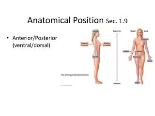
The term Yoga is derived from the Sanskrit root yuj meaning to bind, join, attach and yoke, to direct and concentrate ones attention on, to use and apply. It also means union or communion. It is the true union of our will with the will of God. Yoga is performed through some specific postures called Asana. Among the eight limbs of Yoga, the yogic technique properly begins at the third limb that is the Asana. Asana means a state in which one can remain physically and mentally steady, calm, quiet and comfortable. Patanjali defines Asana as 'Sthirasukhatvam"u009d in Yogasutra which can be translated as STableand pleasurable state of body is called Asana. "Dhanurasana"u009d was described as one of the 32 most important Asana in Gheranda Samhita. The posture is called "Dhanurasana"u009d because in this pose the body resembles a bow with its attached string. The trunk and the thighs represent the bow proper and the hands and legs take the place of a string. In Sanskrit the word Dhanush means a bow. In this article anatomical structures involved in the "Dhanurasana"u009d and how this involvement is beneficial in maintaining the health or in management of any disease is explained. Dr. Somlata Jadoun | Dr. Akanksha Rana | Dr. Sunil Kumar Yadav "An Anatomical Exploration of "Dhanurasana"u009d" Published in International Journal of Trend in Scientific Research and Development (ijtsrd), ISSN: 2456-6470, Volume-4 | Issue-4 , June 2020, URL: https://www.ijtsrd.com/papers/ijtsrd30878.pdf Paper Url :https://www.ijtsrd.com/medicine/other/30878/an-anatomical-exploration-of-u201cdhanurasanau201d/dr-somlata-jadoun<br>

E N D
International Journal of Trend in Scientific Research and Development (IJTSRD) Volume 4 Issue 4, June 2020 Available Online: www.ijtsrd.com e-ISSN: 2456 – 6470 An Anatomical Exploration of “Dhanurasana” Dr. Somlata Jadoun1, Dr. Akanksha Rana1, Dr. Sunil Kumar Yadav2 1PG Scholar, 2Associate Professor, 1,2Department of Sharir Rachana, NIA, Jaipur, Rajasthan, India ABSTRACT The term Yoga is derived from the Sanskrit root yuj meaning to bind, join, attach and yoke, to direct and concentrate one's attention on, to use and apply. It also means union or communion. It is the true union of our will with the will of God. Yoga is performed through some specific postures called Asana. Among the eight limbs of Yoga, the yogic technique properly begins at the third limb that is the Asana. Asana means a state in which one can remain physically and mentally steady, calm, quiet and comfortable. Patanjali defines Asana as ‘Sthirasukhatvam” in Yogasutra which can be translated as STableand pleasurable state of body is called Asana. “Dhanurasana” was described as one of the 32 most important Asana in GherandaSamhita. The posture is called “Dhanurasana” because in this pose the body resembles a bow with its attached string. The trunk and the thighs represent the bow proper and the hands and legs take the place of a string. In Sanskrit the word Dhanush means a bow. In this article anatomical structures involved in the “Dhanurasana” and how this involvement is beneficial in maintaining the health or in management of any disease is explained. KEYWORDS: Yoga, Asana, Anatomy, Joint, Muscle, Dhanurasana How to cite this paper: Dr. Somlata Jadoun | Dr. Akanksha Rana | Dr. Sunil Kumar Yadav "An Anatomical Exploration of “Dhanurasana”" International Journal of Trend in Scientific Research and Development (ijtsrd), ISSN: 2456- 6470, Volume-4 | Issue-4, June 2020, pp.5-11, URL: www.ijtsrd.com/papers/ijtsrd30878.pdf Copyright © 2020 by author(s) and International Journal of Trend in Scientific Research and Development Journal. This is an Open Access article distributed under the terms of the Creative Commons Attribution License (CC (http://creativecommons.org/licenses/by /4.0) Material and Methods - ?Texts related to Yoga-Asana and their commentaries. ?Other print media, online information, journals, magazines etc. Review- According to Gheranda Samhita- ?सा?य?पादौभुिवद?ड?पौकरौचपॄ??े?तपादयु?मम| कॄ?वाधनु?तु?यप?रवित?तािनगधयोगीधनुरासनतत|| (घेर?डसंिहता२/१८) Spreading the legs on the ground, straight like a stick and catching hold of (the toes of) the feet with the hands and making the body bent like a bow, is called by the Yogis the Dhanurasana or Bow posture. According to Hathyogapradipika- पादाङ्गु?ौतुपािण?यां?ही?वा?वणाविध।धनुराकष?नंकुया?दधनुरासनमु?यते॥ (हठयोग?दीिपका1/27) According to Hath Yoga Pradipika, having caught the toes of the feet with both the hands and carried them to the air by drawing the body like a bow, it becomes Dhanurasana. According to Swami Vyas dev ji, Lie down on your belly and take the arms to the back. Bend the knees and also take the feet to the back. Now hold the ankles with the respective hands and stretch them up so that the toes are turned towards the arms. Inhale, retain the breath and raise yourself up. Stretching the arms and legs assume the shape of a bow. Maintain the position as long as you can. Then exhale slowly and return to the normal position.3 Published in IJTSRD30878 BY 4.0) INTRODUCTION “Dhanurasana” was described as one of the 32 important Asana in GherandaSamhita (dated 1650 CE). The Gheranda Samhita is the most encyclopaedic of the three-classic text about Asana. It says that there are 8,400,000 of Asana described by Shiva. The postures are many in number as there are number of species of living creatures in this universe, among them 84 are the best and among these 84, 32 have been found useful for mankind in this world these 32 Asana are mentioned in GherandaSamhita.1 The “Dhanurasana” word comes from the Sanskrit word “Dhanush” meaning A bow and Asana means Posture. Hence, a posture in which the body of practitioner is shaped like a bow with its attached string is called “Dhanurasana”.2 Aim of this study- In this article the important expedition of Asana practitioner about the anatomical structures involved in the Asana and how this involvement is beneficial in maintaining health or in management of any disease and the knowledge of anatomy will also help the Asana practitioners, to avoid injuries. Aim and Objectives - ?To explore the anatomical structures involved in “Dhanurasana.” ?To avoid possibilities of injuries while performing Dhanurasana by understanding the anatomical structures involved in “Dhanurasana”. @ IJTSRD | Unique Paper ID – IJTSRD30878 | Volume – 4 | Issue – 4 | May-June 2020 Page 5
International Journal of Trend in Scientific Research and Development (IJTSRD) @ www.ijtsrd.com eISSN: 2456-6470 Steps for performing Dhanurasana4- ?Lie full length on the floor on the stomach, face downwards. ?Exhale and bend the knees. Stretch the arms back and hold the left foot toe with the left hand and the right foot toe with the right hand. ?Now exhale completely and pull the legs up by raising the knees above the floor, and simultaneously lift the chest off the floor. The arms and hands act like a bow- string to tauten the body like a bent bow. ?Lift up the head and pull it as far back as possible. Do not rest either the ribs or the pelvic bones on the floor. Only the abdomen bears the weight of the body on the floor. ?While raising the legs do not join them at the knees, for then the legs will not be lifted high enough. After the full stretch upwards has been achieved, join together the thighs, the knees and the ankles. Image- Anatomical exploration of “Dhanurasana”- Joint actions5- ?The hip joints are extended, adducted and medially rotated. ?The knees are flexed. ?The ankles are planter flexed. ?Shoulder joints are extended, adducted and internally rotated. ?The elbows are extended. ?Forearms are pronated. ?The spine is extended. Muscles and ligaments involved in “Dhanurasana”- Hip region Hip joints are in extension, adduction and medial rotation in Dhanurasana. Hip extension is the backward movement of the thigh. The extension of hip joint is mainly done by gluteus Maximus and semimembranosus, semitendinosus and long head of biceps femoris. Gluteus Maximus is a primary muscle of the hip extensions. So, the flexor of the hip joint will get stretch. The flexors of hip joint are Sartorius, vastus lateralis and vastus medialis and vastus intermedius. The nerve supply of these muscles is femoral nerve. The Adduction of hip joint is done by adductors longus, brevis and magnus and it is assisted by the pectineus and gracilis. During adduction of the hip joint gluteus medius and minimus stretched. The medial rotation of hip joint is done by Tensor fasciae latae and the anterior fibres of the glutei medius and minimus. So, the primary abductors gluteus medius, gluteus minimus and tensor fasciae latae and the lateral rotators of hip joint obturator internus and quadratus femoris are stretched. @ IJTSRD | Unique Paper ID – IJTSRD30878 | Volume – 4 | Issue – 4 | May-June 2020 Page 6
International Journal of Trend in Scientific Research and Development (IJTSRD) @ www.ijtsrd.com eISSN: 2456-6470 Table1. Muscles performing hip extension, adduction and medial rotation in Dhanurasana Muscle Position Gluteus maximus Gluteal region Semitendinosus Posterior compartment of thigh Semimembranosus Posterior compartment of thigh Long head of biceps femoris Posterior compartment of thigh Adductor longus Medial compartment of thigh Adductor brevis Medial compartment of thigh Gracilis Medial compartment of thigh Pectineus Medial compartment of thigh Tensor fasciae latae Gluteal region Nerve supply Inferior gluteal nerve (L5-S2) Sciatic nerve (L5-S2) Sciatic nerve (L5-S2) Tibial part of sciatic nerve Obturator nerve (L2-L4) Obturator nerve (L2-L4) Obturator nerve (L2-L4) Femoral nerve (L2, L3) Superior gluteal nerve (L4-S1) Table2. Muscles which are stretched at hip joint in Dhanurasana Muscles Position Sartorius Anterior compartment of thigh Vastus lateralis Anterior compartment of thigh Vastus medialis Anterior compartment of thigh Vastus intermedialis Anterior compartment of thigh Gluteus Medius Gluteal region Gluteus minimus Gluteal region Quadratus femoris Gluteal region Obturator internus Gluteal region Nerve supply femoral nerve (L2, L3) femoral nerve (L2, L3) femoral nerve (L2, L3) femoral nerve (L2, L3) Superior Gluteal nerve (L4, L5, S1) Superior Gluteal nerve (L4, L5, S1) Superior Gluteal nerve (L4, L5, S1) Superior Gluteal nerve (L4, L5, S1) Knee region Knee joint is flexed. The flexion of the knee joint is mainly done by semimembranosus, semitendinosus and biceps femoris and it is assisted by the gracilis, popliteus and sartorius. The extensor compartment or anterior compartment of the thigh will get stretched in Dhanurasana. This compartment consists of quadriceps femoris which includes rectus femoris, vastus lateralis, medialis and intermedius. Table3. Muscles performing flexion of the knee in Dhanurasana Muscle Position Long head by tibial part and short head by common peroneal part of sciatic nerve (L5, S1, S2) Semimembranosus Back of thigh Semitendinosus Back of thigh Table4. Muscles which are stretched at knee joint in Dhanurasana. Muscle Position Vastus medialis Anterior compartment of thigh Femoral nerve (L2-L4) Vastus intermedius Anterior compartment of thigh Femoral nerve (L2-L4) Vastus lateralis Anterior compartment of thigh Femoral nerve (L2-L4) Rectus femoris Anterior compartment of thigh Femoral nerve (L2-L4) Ligaments of knee joint Knee joint is flexed. In this position the maximum pressure is on the following ligaments. ?Medial and lateral meniscus. ?Posterior cruciate ligament. Ankle region The ankle is plantarflexed and the foot is inverted in Dhanurasana. Ankle plantar flexion is performed by group of muscles in the posterior compartment of leg and the extrinsic and intrinsic muscles of the toe joint. Gastrocnemius, soleus, tibialis posterior, plantaris, Peroneus longus and brevis. The extrinsic muscles of the toe joint which assist plantar flexion are flexor hallucis longus, and flexor digitorum longus. The intrinsic flexors of toe joints are flexor digitorum brevis, flexor hallucis brevis, quadratus plantae, lumbricals and flexor digiti minimi brevis. The extensor muscles of the anterior compartment of leg are stretched when the ankle is plantarflexed. This includes extensor digitorum longus, extensor hallucis longus, tibialis anterior and peroneus tertius. During the inverted position of the foot evertor muscles of the lateral compartment are stretched. Peroneus longus and brevis belong to lateral compartment of leg. The muscles of dorsum of foot are also stretched. Extensor digitorum brevis and extensor hallucis brevis belongs to the dorsum of foot. Nerve supply Biceps femoris Back of thigh Tibial part of sciatic nerve (L5, S1, S2) Tibial part of sciatic nerve (L5, S1, S2) Nerve supply @ IJTSRD | Unique Paper ID – IJTSRD30878 | Volume – 4 | Issue – 4 | May-June 2020 Page 7
International Journal of Trend in Scientific Research and Development (IJTSRD) @ www.ijtsrd.com eISSN: 2456-6470 Table5. Muscles performing ankle planter flexion in Dhanurasana Muscle Position Gastrocnemius Posterior compartment of leg Soleus Posterior compartment of leg Plantaris Posterior compartment of leg Tibialis posterior Posterior compartment of leg Flexor hallucis longus Posterior compartment of leg Flexor digitorum longus Posterior compartment of leg Flexor digitorum brevis Plantar Lumbricals Plantar Quadratus plantae Plantar Flexor hallucis brevis Plantar Flexor digiti minimi Plantar Nerve supply Tibial nerve (S1, S2) Tibial nerve (S1, S2) Tibial nerve (S1, S2) Tibial nerve (L4, L5) Tibial nerve (L5, S1, S2) Tibial nerve (L5, S1, S2) Medial plantar nerve (S1, S2) Medial and lateral plantar nerve (S2, S3) Lateral plantar nerve (S1, S2) Medial plantar nerve (S1, S2) Lateral plantar nerve (S1, S2) Table6. Muscles which are stretched at ankle joint in Dhanurasana Position Tibialis anterior Anterior compartment of leg Extensor digitorum longus Anterior compartment of leg Extensor hallucis longus Anterior compartment of leg Peroneus tertius Anterior compartment of leg Peroneus longus Lateral compartment of leg Peroneus brevis Lateral compartment of leg Extensor digitorum brevis Dorsum of foot Extensor hallucis brevis Dorsum of foot Muscle Nerve supply Deep peroneal nerve (L4-S2) Deep peroneal nerve (L4-S2) Deep peroneal nerve (L4-S2) Deep peroneal nerve (L4-S2) Superficial peroneal nerve (L5, S1, S2) Superficial peroneal nerve (L5, S1, S2) Terminal branches of deep peroneal nerve (S1, S2) Terminal branches of deep peroneal nerve (S1, S2) Ligaments of Ankle joint Ankle is in planter flexion. In this position the maximum pressure is on the following ligaments. ?Anterior talofibular ligaments (ATFL) ?Posterior talofibular ligaments (PTFL) ?Calcaneofibular ligament Shoulder region Shoulder joint is extended, adducted and internally rotated in the pose of Dhanurasana. Shoulder girdle is depressed. The Extension of shoulder joint is caused by Posterior fibres of deltoid, Latissimus dorsi and assisted by the Teres major, Long head of triceps, Sternocostal head of the pectoralis major. The muscles acting as antagonists for this action are clavicular head of pectoralis major, anterior fibres of deltoid, coracobrachialis and short head of biceps. They are stretched when the shoulder joint is extended. Adduction of shoulder joint is principally done by the Pectoralis major, Latissimus dorsi, Short head of biceps, Long head of triceps, and it is assisted by coracobrachialis and teres major which is antagonised by deltoid and supraspinatus. Medial rotation of shoulder joint is done by the pectoralis major, anterior fibres of deltoid, latissimus dorsi, teres major and subscapularis. The muscles acting as antagonists for this action are infraspinatus, teres minor and posterior fibres of deltoid. Pectoralis minor muscle helps in depression and anterior tilt of scapula and is stretched by the posterior pull of scapula. Deltoid muscle has large range of action on shoulder joint. It helps in the abduction of shoulder joint, the anterior fibers in flexion and posterior fibres in external rotation. Supraspinatus along with deltoid helps in abduction of shoulder joint. Infraspinatus and teres minor are external rotators along with the posterior fibers of deltoid. Table7. Muscles performing shoulder joint extension, adduction and internal rotation. Muscle Position Deltoid Scapular Latissimus dorsi Back Pectoralis major Thorax Biceps Anterior compartment of Arm Coracobrachialis Anterior compartment of Arm Triceps Posterior compartment of arm Teres major shoulder Subscapularis shoulder Nerve supply Axillary nerve(C5-C6) Thoracodorsal nerve (C6-C8) Medial and lateral pectoral nerve (C5-T1) Musculocutaneousnerve(C5-C7) Musculocutaneousnerve(C5-C7) Radial nerve (C6-C8) Lower subscapular nerve (C5, C6) Upper and lower subscapular nerve (C5, C6) @ IJTSRD | Unique Paper ID – IJTSRD30878 | Volume – 4 | Issue – 4 | May-June 2020 Page 8
International Journal of Trend in Scientific Research and Development (IJTSRD) @ www.ijtsrd.com eISSN: 2456-6470 Table8. Muscles which are stretched at shoulder joint in Dhanurasana. Muscle Position Supraspinatus Scapular Infraspinatus Scapular Teres minor Scapular Trapezius Scapular Levator scapulae Scapular Serratus anterior Scapular Pectoralis minor Thorax Medial and lateral pectoral nerve (C5-T1) Nerve supply Suprascapular nerve (C5, C6) Suprascapular nerve (C5, C6) Axillary nerve (C5, C6) Accessory nerve Dorsal scapular nerve (C4, C5) Long thoracic nerve (C5-C7) Scapula elevation Elevation of scapula is caused by the Levator scapulae it’s also pulled down in this Asana. Serratus anterior muscle helps in protraction and upward rotation of scapula. This muscle is stretched by the downward pull of scapula. Elbow region Elbow extended and Forearm pronated in Dhanurasana. Extension of elbow joint is completed by the triceps brachii and anconeus, and pronation of radioulnar joint is caused by the pronator teres and pronator quadratus. In Dhanurasana the upper limb is kept straight and the elbow is extended. The forearm is in pronated position. To maintain the extension of elbow joint the triceps brachii is actively contracted. Table9. Muscles performing elbow joint extension in Dhanurasana. Muscle Position Triceps brachii Posterior compartment of arm Anconeus Posterior compartment of forearm Radial nerve (C5-T1) Table10. Muscles which are stretched at elbow joint in Dhanurasana. Muscle Position Brachialis Anterior compartment of arm Brachioradialis Posterior compartment of forearm The Spine Similarly, to Bhujangasana in Dhanurasana also the spine is completely extended. All extensors of the back along with external oblique and transverse abdominus are contracted in Dhanurasana. These include the erector spinae muscles, transvers spinalis muscles, quadratus lumborum and Levator costarum. The thoracic and lumbar spines are in extension. The muscles of anterior abdominal wall help in the Extension of trunk. These includes rectus abdominus, external oblique abdominus and internal oblique abdominus. Table11. Muscles performing spine extension in Dhanurasana. Nerve supply Radial nerve (C5-T1) Nerve supply musculocutaneous nerve (C5, C6) Radial nerve (C5-T1) Muscle Position Nerve supply Lateral branches of the Dorsal rami of the cervical, thoracic and lumbar spinal nerves. Lateral branches of the Dorsal rami of the cervical, thoracic and lumbar spinal nerves. Lateral branches of the Dorsal rami of the cervical, thoracic and lumbar spinal nerves. Lateral branches of the Dorsal rami of the cervical, thoracic and lumbar spinal nerves. Medial branches of the dorsal rami of the appropriate spinal nerves. Medial branches of the dorsal rami of the appropriate spinal nerves. Medial branches of the dorsal rami of the appropriate spinal nerves. Erector spinae Back Iliocostalis Back Longissimus Back Spinalis Back Semispinalis Back Multifidi Back Rotatores Back Levator costarum Back Dorsal rami C8-T11 (Intercostal nerves) Ventral rami of the twelfth thoracic and upper three or four lumbar spinal nerves. Quadratus lumborum Posterior abdominal wall @ IJTSRD | Unique Paper ID – IJTSRD30878 | Volume – 4 | Issue – 4 | May-June 2020 Page 9
International Journal of Trend in Scientific Research and Development (IJTSRD) @ www.ijtsrd.com eISSN: 2456-6470 Table12. Muscles which are stretched at trunk in Dhanurasana. Position Rectus abdominis Anterior abdominal wall External oblique Anterior abdominal wall Internal oblique Anterior abdominal wall Ventral rami of the lower six thoracic and first lumbar spinal nerves. Muscle Nerve supply Ventral rami of the lower six or seven thoracic spinal nerves. Ventral rami of the lower six thoracic spinal nerves. Cervical region Cervical spine is extended. In this position the extensors of cervical region are contracted. Trapezius, splenius capitis, splenius cervicis, semispinalis capitis and longissimus Capitis helps to extend the head and are contracted in this case. The suboccipital muscles are Rectus capitis posterior major, Rectus capitis posterior minor, Obliquus capitis inferior and Obliquus capitis superior are involved in extension of the head at the Atlanto-occipital joints and rotation of the head and atlas on the axis. These are also stretched in this Asana. Thoracic spine It is extended. The superior thoracic vertebrae glide inferior and posterior. Iliocostalis thoracis, Longissimus thoracis, Spinalis thoracis, Multifidus, Semispinalis thoracis are active contracted in Dhanurasana. Lumbar spine It is extended. Extrinsic back muscles, in the superficial layers Latissimus Dorsi, Levator Scapulae, Rhomboids, trapezius contracts while extension of the lumbar region. Intrinsic muscles help in extension of lumbar spine, Iliocostalis, Longissimus, Spinalis, Semispinalis contracts while performing the Dhanurasana. Anterior abdominal wall muscles stretched in Dhanurasana. Wrist and Hand The hand holds the big toe and the metacarpophalangeal (MCP) and interphalangeal (IP) are flexed to grasp the toe. Primarily flexion of the IP and MCP joints is from the flexor digitorum profundus. The flexor digitorum superficialis assists when increased forces are required. Flexor pollicis brevis and opponens pollicis flex the thumb. Interossei and lumbrical muscles flex the MCP joints. Flexor digitorum superficialis and profundus flex the IP joints of the second through fifth digits. Because the tendons of these muscles pass on the palmar side of the wrist and the MCP joints, they also tend to flex these joints. In using the hand for grasping, flexion of the MCP joints is necessary for the hand to assume the shape of the object grasped or to properly shape the hand for its desired use. Table13. Muscles performing flexion of wrist and hand in Dhanurasana. Muscle Position Flexor digitorum superficialis Anterior compartment of forearm Nerve supply Median nerve Medial part-ulnar nerve. Lateral part-median nerve (C8, T1) Median nerve (C7, C8) Median nerve Deep branch of ulnar nerve Median nerve and ulnar nerve Flexor digitorum profundus Anterior compartment of forearm Flexor pollicis longus Flexor pollicis brevis Interossei Lumbricals Anterior compartment of forearm Hand Hand Hand Benefits of “Dhanurasana”- It exerts great pressure on spine and stomach and thus strengthens the vertebral bones; spine becomes elastic; releases gas of the stomach; relieves constipation and dyspepsia. Muscles and nerves of shoulders, arms, hands, thighs and feet are benefitted.6 In this posture the spine is stretched back. Elderly people do not normally do this, so their spines get rigid. This Asana brings back elasticity to the spine and tones the abdominal organs. In my experience, persons suffering from slipped discs have obtained relief by the regular practice of Dhanurasana and Shalabhasanawithout being forced to rest or to undergo surgical treatment.7 Discussion The basic joint position in Dhanurasana is extension of spine. Extension, medial rotation, adduction of Hip joint, Flexion of knee joint and extension, medial rotation and adduction of shoulder joint. Dhanurasana is very important for stimulating the solar plexus. It regulates the digestive, eliminatory and reproductive organs. While performing the Dhanurasana anterior abdominal wall muscles get maximum stretch. The abdominal wall is innervated by intercostal nerves (arising from ilioinguinal/iliohypogastric nerves (arising from L1). Dhanurasana stimulates the nerves of abdominal wall and affects the sympathetic and parasympathetic functions of organs. It massages the liver and pancreas and thus very useful for yogic management of diabetes. The kidneys are stimulated and the whole alimentary canal is toned. By lying on the diaphragm with the shoulder is extended, the heart is given a gentle massage and, because the thorax is fully expanded in this posture. Dhanurasana improves the blood circulation around muscles and tendons and organs but also improves the muscle tone and greater elasticity of ligaments. The blood pressure, heart rate if increased, comes to the normal or remains within normal range without putting extra burden on cardio-respiratory system. T6 to T12) and @ IJTSRD | Unique Paper ID – IJTSRD30878 | Volume – 4 | Issue – 4 | May-June 2020 Page 10
International Journal of Trend in Scientific Research and Development (IJTSRD) @ www.ijtsrd.com eISSN: 2456-6470 Dhanurasana is useful in the treatment of various chest ailments. It stimulates and regulates the endocrine glands, particularly the thyroid and adrenal glands, and it induces production of cortisone. The extension of spine adjusts the vertebral column, straightening a hunched back and drooping shoulders. It is also recommended for treating certain types of rheumatism. Dhanurasana helps to regulate the menstrual cycle and also to correct female infertility, if the cause is not due to deformity of the reproductive organs themselves. Conclusion In Dhanurasana, having caught the toes of the feet with both the hands and carried them to the air by drawing the body like a bow. In this Asana shoulder joint, hip joint is under more stress. The muscles of pectoral region, anterior compartment of arm, anterior abdominal wall and anterior compartment of thigh stretched. The thorax is fully expanded in this posture. Dhanurasana improves the blood circulation around muscles and tendons and organs. Stimulates and regulates the endocrine glands, particularly the thyroid and adrenal glands Sacroiliac joint, hip joint and knee joint limits this posture there are chances of injuries to ligaments in these joints. References [1]The GherandaSamhita translated in English by Rai Bahadur Srisa Chandra Vasu; published by Sri Satguru Publications; New Delhi; Reprint 1979 [2]Iyengar, B. K. S. (1979). Light on Yoga,Yoga Dipika (revised ed),foreword by Yehudi Menuhin Schocken Books New York pg no. 282 [3]Dev SV. First Steps to Higher Yoga. First Edit. Yoga Niketan trust; 1970. Page 140 [4]Saraswati SS. Asana Pranayama Mudra Bandha. Fourth Edi. Munger: Yoga Publication Trust;2009. Page 211 [5]Leslie Kaminoff, Yoga Anatomy, illustrated by Sharon Ellis, Human kinetics, page no. 190 [6]Iyengar BKS. Light on Yoga. revised ed. Schocken Books New York; 1979. [7]Iyengar BKS. Light on Yoga. revised ed. Schocken Books New York; 1979 @ IJTSRD | Unique Paper ID – IJTSRD30878 | Volume – 4 | Issue – 4 | May-June 2020 Page 11