Download
1 / 33
330 likes | 437 Views
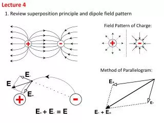
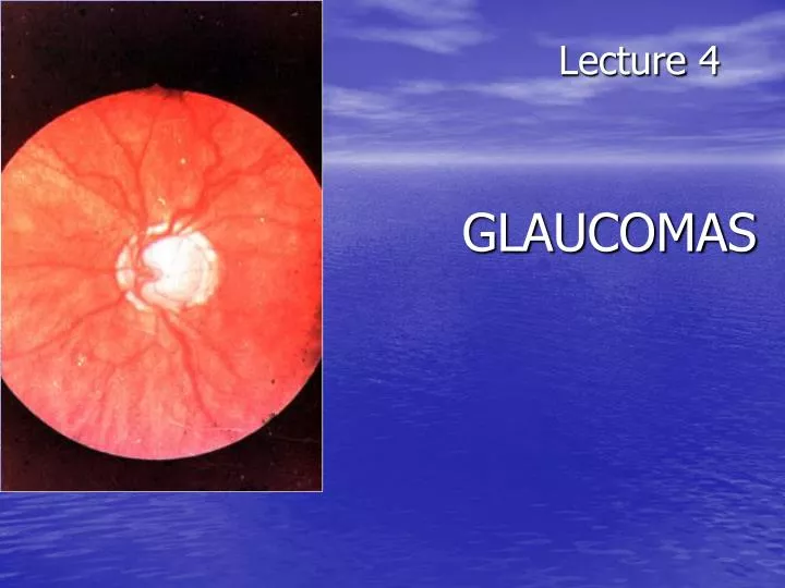
Lecture 4 GLAUCOMAS. The outflow pathways of aqueous humor : Main : posterior chamber - p u pil - anterior chamber - trabecular meshwork - Schlemm’s canal (scleral sinus)- vorticose veins – scleral venous plexus. Additional : 2. Perivascular spaces of iris.

E N D
The outflow pathways of aqueous humor: • Main: posterior chamber - pupil - anterior chamber - trabecular meshwork - Schlemm’s canal (scleral sinus)- vorticose veins – scleral venous plexus. • Additional: • 2. Perivascular spaces of iris. • 3. Suprachoroidal space - perivascular spaces – through sclera into the tenon’ s space. • 4. Perivascular spaces of central retinal vessels. • The IOP is maintained by a balance between aqueous inflow and outflow &usually measures between • 16-26 mm Hg (using tonometr of Maklakov) & • 10-20mm Hg (using tonometr of Goldman)
Not every increasing of IOP is glaucoma. It may be ocular hypertension, caused, for example, by using corticosteroids, intoxication or climax. Typical for ocular hypertension are: • absence of structural and functional changes; • lasting existence without complaints; • symmetrical increasing of IOP. • So, ocular hypertension is a symptom, glaucoma is a syndrome. • Glaucoma is such increasing of IOP, which is accompanied by specific visual defects (constriction of nasal visual field, Bjerrum’s scotoma) and specific optic disc changes (dislocation of vessels, increased cuppingetc.)
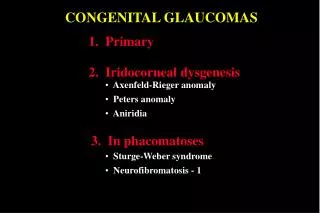
Congenital glaucoma (or hydrophtalmos) is caused byabnormal development of eye drainage system. The accumulation of aqueous in the eye due to elasticity of baby’s external coat causes the increasing of eye size. • There are 2 clinical forms: • Hydrophtalmos without stasis (megalocornea, stretching out of limbus, deep anterior chamber, increased eye, loss of vision, increased IOP, typical changes of optic nerve). • II. Hydrophtalmos with stasis (all above mentioned signs + photophobia, blepharospasmus, mixt injection, corneal oedema, which is reliefed by 40 % glucosae). • There are 4 stages: • I. Early – D of cornea 12,0-12,5 mm, anterior-posterior distance of the eye is increased on 1,5-2,0 mm, N fundus. • II. Advanced - D of cornea 13,0-14,0 mm, anterior-posterior distance of the eye is increased on 3,0-4,0, glaucomatous cupping of optic disc ophthalmoscopically. • III. Far advanced - D of cornea is more then 14,0 mm, anterior-posterior distance of the eye is more then 30,0 mm, atrophy of optic disc ophthalmoscopically. • IV. Terminal (or buftalm) – full blidness, scleral staphyloma.
Methods of diagnostic of congenital glaucoma: • General examination, especially of cornea & limbus • Biomicroscopy or focal lighting • Keratometry • Tonometry • Ultrasound biometry • Ophthalmoscopy • Methods of treatment of congenital glaucoma: • Only surgical. Immediatly! • Goniotomy • Sinusotrabeculectomy • Enucleation in buftalmos
Stages of primary glaucoma (according to visual functions defects): I – visual field is consticted less then 10 degrees, physiological cupping is increased. II - visual field is consticted more then 10 degrees, edge excavation. III – tube visual field (15 degrees from the point of fixation), edge excavation. IV – visual field or visual acuity is zero, atrophy of optic disc. Depending on IOP (using tonometr of Maklakov) glaucoma is subdivided: A (compensated) – IOP is less then 27 mm Hg. B (subcompancated) – IOP is 28-32 mm Hg. C (decompancated) – IOP is 33 mm Hg and more. According to dynamics of visual functions during 6 month: stabile & nonstabile – constriction of visual field on 10 degree and more; in tube vision – on 2-3 degrees and more; increasing of scotomas size; increasing of size of optic disc cupping
Open-angle glaucoma • Pathogenesis – constriction or closing of openings in trabeculae & Schlemm’s canal as a result of endocrine, vascular or general diseases such as atherosclerosis, artery hypertension, diabetus mellitus etc. • Clinical features:usually asymptomatic until significant loss of visual field has occured; • the eye looks usual, only dystrophic iris changes may be revealed biomicroscopically; • open anterior chamber angle on gonioscopy, may be excess pigmentation of trabeculae; • & typical for glaucoma signs (elevated IOP+visual field loss,first of its nasal part +optic nerve damage). • Methods of investigation: • Functional – visometry, perimetry, campimetry, adaptometry. • B. Objective – general examination, focal lighting, biomicroscopy, gonioscopy, ophthalmoscopy, tonometry.
Close-angle glaucoma Pathogenesis –the closing (blockade) of anterior chamber angleby iris root as a result of excess anterior position of lens or functional pupil blockade (not organic, i.e. occlusio or seclusio pupille) due to excess near location of lens & iris. Clinical features:complaints for clouding of vision, haloes around lights in the morning, headache, pain in the eye etc.; frequent change of eye refraction & glasses prescribtion; sometimes begins from acute attack; signs of venous stasis – dilated scleral veins; flat anterior chamber & iris bombee biomicroscopically; narrow or close anterior chamber angle on gonioscopy; & typical for glaucoma signs (elevated IOP+visual field loss,first of its nasal part +optic nerve damage). Methods of investigation: A. Functional – visometry, perimetry, campimetry, adaptonetry. B. Objective – general examination, focal lighting, biomicroscopy, gonioscopy, ophthalmoscopy, tonometry.
Medical treatment of chronic glaucoma: • 1. Local hypotensive therapy. The antiglaucomatous drops are divided into two main groups: • I. which improve outcome of aqeous humour • Cholinomimetics - 1 % pilocarpini, carbachol; • α, Β – adrenomimetics – dipinefrini, epinefrini; • Analogs of prostaglandins F 2 α (which stimulate the uveo-scleral outflow) – latanoprost (xalatan), travoprost (travatan) • II. which reduce production of aqeous humour • Central agonists of α2- adrenoreceptors - klonidini; • B-adrenoblockers: nonselective - timololi, arutimoli, & selective - betoptic; • Carbonic anhydrase inhibitors – Azopt. 2. Vasodilatators – acidi nicotinici, cavintoni, trentali, halidori etc. 3. Nootrops – piracetami, nootropili, etc. 4. Stimulators of nerve conductivity – proserini. 5. Tissue therapy, vitamins. • Laser treatment of chronic glaucoma: • Laser peripheral iridotomies in primary angle-closure glaucoma; • Laser trabeculoplasty in primary open-angle glaucoma. • Surgery of chronic glaucoma: • Filtration surgery in primary open-angle glaucoma, e.g. trabeculectomy. • In primary angle-closure glaucoma radical surgery – phacoemulsification of cataract with IOL implantation; palliative surgery – iridectomy.
Nonpenetreting filtration surgery:canaloplasty • Figure 1 (left). Introduction of the microcatheter into Schlemm's canal • Figure 2 (right). A 10-0 polypropylene suture being tied around the end of themicrocatheter
Nonpenetreting filtration surgery:viscocanalostomy The initial steps of viscocanalostomy are similar to those of trabeculectomy. Specifically, the surgeon creates a one-half– to two-thirds–depth superficial scleral flap, within the bed of which a deep scleral flap is made. The deep dissection begins 4 to 5 mm posterior to the limbus and advances toward the limbus in a tissue plane just above the suprachoroidal space. As the dissection advances anteriorly, the roof of Schlemm’s canal is removed. The surgeon then cannulates Schlemm’s canal and injects a bolus of viscoelastic material into each of the canal’s cut ends (as in the picture). This viscodissection is intended to dilate the canal and facilitate the subsequent drainage of aqueous.
Emergency in acute close-angle glaucoma: • instillation of miotics (pilocarpini 1 or 2 %) every 15 minutes during first hour, every 30 minutes during next hour, then 4 times a day; • analgetics (promedoli 2 % 1,0 ml s/cutaneous); • diuretics (Diacarbi 0,5 or Hipothiasidi 0,1 per os, Lasix 1 % 2,0 ml i/m) • If the attack of acute close-angle glaucoma doesn’t disappear during 12-24 hours, • antiglaucomatous surgery is indicated.
Suspicion of glaucoma may be in such cases: • IOP is 27 mm Hg and more (using tonometr of Maklakov) and 21 mm and more (using tonometr of Goldman); • complaints for clouding of vision, haloes around lights in the morning; • iris bombee, less depth of anterior chamber; • typical changes of optic disc; • the difference in right and left eye IOP is more then 5 mm Hg. • All patients with suspicion of glaucoma must be observed in details in clinics. This diagnosis can exist only one year. • Methods of investigation: • A. Functional – visometry, perimetry, adaptonetry, campimetry. • B. Objective – general examination, focal lighting, biomicroscopy, gonioscopy, ophthalmoscopy, tonometry. • C. Necessary additional – • diurnal tonometry, tonography, elastotonometry, provocative test.
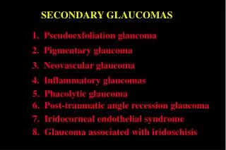
Secondary glaucoma is complication or outcome of some other eye diseases. It may be: 1. Uveal glaucoma – as a result of pupil occlusion. Management – treatment of uveitis. In deep anterior chamber– mydriatics. In flat anterior chamber – miotics. 2. Phacogenic – prodused by immature cataract or lens dislocation into the anterior chamber. Management – cataract surgery. 3. Phacolytic - prodused by hypermature cataract. Management – cataract surgery. 4. Vascular glaucoma as a result of central vein occlusion or neovascularization in diabetus mellitus. Management – treatment of main disease. 5. Posttraumatic as a result of burns, penetrating or blunt injury of eyeball. Management – miotics. 6. Neoplastic – as a result of intraocular tumours. Management–surgery (enucleation).