Download
1 / 15
470 likes | 2.23k Views
SECONDARY GLAUCOMAS. 1. Pseudoexfoliation glaucoma. 2. Pigmentary glaucoma. 3. Neovascular glaucoma. 4. Inflammatory glaucomas. 5. Phacolytic glaucoma. 6. Post-traumatic angle recession glaucoma. 7. Iridocorneal endothelial syndrome. 8. Glaucoma associated with iridoschisis.

E N D
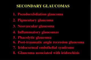
SECONDARY GLAUCOMAS 1. Pseudoexfoliation glaucoma 2. Pigmentary glaucoma 3. Neovascular glaucoma 4. Inflammatory glaucomas 5. Phacolytic glaucoma 6. Post-traumatic angle recession glaucoma 7. Iridocorneal endothelial syndrome 8. Glaucoma associated with iridoschisis
Pseudoexfoliation glaucoma • Secondary trabecular block open-angle glaucoma • Affects elderly, unilateral in 60% • Prognosis less good than in POAG Iris sphincter atrophy Gonioscopy Pseudoexfoliative material Central disc with peripheral band On retroillumination Trabecular hyperpigmentation - may extend anteriorly (Sampaolesi line)
Pigmentary glaucoma • Bilateral trabecular block open-angle glaucoma • Typically affects young myopic males • Increased incidence of lattice degeneration Krukenberg spindle and very deep anterior chamber Fine pigment granules on anterior iris surface Trabecular hyperpigmentation Mid-peripheral iris atrophy
Causes of neovascular glaucoma • Common, secondary angle-closure glaucoma without pupil block • Caused by rubeosis iridis associated with chronic, diffuse retinal ischaemia Long-standing diabetes (common) Ischaemic central retinal vein occlusion (most common) Central retinal artery occlusion (uncommon) Carotid obstructive disease (uncommon)
Signs of advanced neovascular glaucoma Severe rubeosis iridis Severely reduced visual acuity, congestion and pain Synechial angle closure Distortion of pupil and ectropion uveae
Treatment options of neovascular glaucoma Topical • Atropine and steroids to decrease inflammation • Beta-bockers Panretinal photocoagulation - in early cases Artificial filtering devices - in very advanced cases Cyclodestructive procedures - to relieve pain Retrobulbar alcohol injection - to relieve pain
Inflammatory glaucomas Angle-closure with pupil block • Caused by seclusio pupillae • Anterior chamber is shallow
Inflammatory glaucomas Angle-closure without pupil block • Caused by progressive synechial angle closure • Anterior chamber is deep
Phacolytic glaucoma Pathogenesis Signs Treatment • Control IOP medically • Deep anterior chamber • Remove cataract • Floating white particles
Post-traumatic angle recession glaucoma Pathogenesis Signs Irregular widening of ciliary body band Blunt traumatic damage to trabecular meshwork
Classification of Iridocorneal Endothelial Syndrome • Proliferation of abnormal corneal endothelial cells • Typically affects young to middle aged women • Three syndromes with certain overlap 1. Progressive iris atrophy • Iris atrophy in 100% 2. Iris naevus (Cogan-Reese) syndrome • Iris atrophy in 50% 3. Chandler syndrome • Iris atrophy in 40% • Corneal changes predominate
Progressive iris atrophy Progressive stromal iris atrophy Displacement of pupil towards PAS Broad-based PAS
Iris naevus (Cogan-Reese) syndrome Diffuse iris naevus Pedunculated iris nodules
Chandler syndrome Later oedema which may cause halos Initially ‘hammer-silver’ endothelial changes
Glaucoma associated with iridoschisis • Rare, affects elderly, often bilateral • Underlying, angle-closure glaucoma in about 90% Shallow anterior chamber Iridoschisis - usually inferior