Understanding the Human Eye: Anatomy and Vision Theories
Explore the intricate workings of the human eye, from its anatomy—including the cornea, lens, and retina—to the complex processes behind vision and color perception. Learn about key structures like the optic nerve and pupil, and discover essential concepts like light adaptation, afterimages, and the significance of rod and cone cells. Engage with interactive quizzes to test your knowledge and delve into fascinating color vision theories, including trichromatic and opponent-process theories. Enhance your appreciation for how we perceive the world around us!

Understanding the Human Eye: Anatomy and Vision Theories
E N D
Presentation Transcript
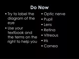
Do Now Optic nerve Pupil Lens Retina Vitreous Iris Cornea • Try to label the diagram of the eye • Use your textbook and the terms on the right to help you
Vision • The Visual System • Color Vision and Theories
Vision Test!!! • First stare continuously at the center of the upper square for about 20 seconds, then look at the dot in the lower square. • Within a moment, a gray-and-white afterimage should appear inside the lower square
The Visual SystemStructures and Functions • Cornea • Clear protective coating that covers the front of the eye • Where light enters the eye • Pupil • Opening in the center of the eye to let light pass through • Iris • Colored part of the eye • Contract in bright light (protects eye) • Expand in dim light (let in extra light) Iris Cornea Pupil
The Visual SystemStructures and Functions • Lens • Transparent, inside pupil • Focuses light on retina • Changes shape to see near vs. far • Retina • Inner surface in back of eyeball • Contains receptor cells • Very center is called the fovea Vitreous Iris Retina Cornea Optic Nerve Lens Pupil
Find Your Blind Spot Place on the retina where the optic nerve enters the retina and where there are no receptor cells
Location = retina Sensitive to electromagnetic energy, primarily light Rods Respond to intensities of light/dark Night vision - sensitive to light About 120 million Cones Responsible for seeing colors Found in fovea About 8 million Bipolar cells Specialized neurons connected to receptors The Visual SystemReceptor Cells
Do Now • Fill out Receptor Cell info on packet page #1
The Visual SystemAdaptation • Dark adaptation • Process by which rods and cones become more sensitive to darkness • Not enough light to activate cones • CANNOT SEE COLOR • Light adaptation • Process by which sensitivity of rods and cones decreases in bright light
This is called afterimage Sense experience that occurs after a visual stimulus has been removed
Pop Quiz!!! What do you see with? YOUR BRAIN
Color VisionProperties of Color • Hues • Aspects of color that correspond to general color names like red, green, and blue • Saturation • Vividness or richness of a hue • Forrest green, olive green, pale green, neon green, etc. • Brightness • Based on the nearness of a color to white as opposed to black • Is the color more white or black?
Color VisionTheories • Primary colors for light mixtures: red, green, blue • Additive color mixing • Mixing light of different wavelengths to create new hue • Subtractive color mixing • Mixing pigments • Concerned with what colors are absorbed and reflected • Trichromatic theory • Perception of all colors is based on three color receptors (red, green, blue) in the retina • Opponent-process theory • Color receptors come in pairs (yellow-blue, red-green, black-white) • Trichromatic = normal vision • Color blindness…dichromat and monochromat
Color VisionFun Facts!!! • Color is in the eye of the beholder • Humans and most primates are trichromats • Rodents and owls (nocturnal animals) are monochromats • Bees can see ultraviolet light