Download

1 / 121
1.21k likes | 1.29k Views
Explore the macroscopic anatomy of the breast, including the conventional partition into 4 quadrants, areola, axillary part, and inframammary fold. Understand the functional structure with glandular, connective, and fatty tissue compositions that vary with age, nutrition, and physiological status. Learn about areola characteristics, male breast rudimentary features, breast development changes, aging effects, blood vessels/lymphatics anatomy, and congenital malformations.

E N D
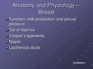
Macroscopic anatomy • Conventional partition • 4 quadrants • Areola • Axillary part • Inframamary fold
Functional structure of the breast • Please review physiology
3 ESENTIAL COMPONENTS • Glandular tissue • Connective tissue • Fatty tissue • Their proportions varies significantly with • Age • Nutrition status • Pregnancy/Post-partum/Lactation
1.Glandular tissue Produces milk as final product 15-20 lobules completely separated, with radial disposition around the nipple Galactofore ducts – nipple (small dilated area in the areola, opening in the nipple) Microscopic structure
2 Conective tissue Creates structure/ support Included are the suspensory ligaments Connects the breast to the skin and fascia of the pectoralis major Microscopic structure
3 Fatty tissue Participates to the structure of the breast Anterior, posterior and within the lobules Varies according to diet and can produce major variations in the volume of the breast Microscopic structure
Areola • Specialized skin adapted for lactation • Nipple in the middle • Sebaceous glands visible on the surface (Montgomery) • Contains smooth muscles which participate in milk evacuation during lactation • Highly innervated area
Male breast • Rudimentary, areola and nipple • Glandular tissue + fatty tissue are – most often- rudimentary or absent. Normal <2cm
Breast development • Significant changes in volume and shape according to • Age • Physiologic status • In adolescence structure becomes nodular and the volume and structure changes during menstrual cycle.
Aging • Volume diminishes • Changes in structural proportions – glandular tissue diminishes and is replaced by fat • Changes in position due to loss in the elasticity of suspensor structures • Loss of axillary hair • Sclerosing of ducts
Blood vessels and lymphatics • Please review anatomy
Axilary lymph nodes • Please review anatomy
Congenital malformations • Breast develops from the mammary folds (ectodermic epithelium) on a virtual line (axilla – inguinal ligament) • On this line mammary buds develop and regress spontaneously • At birth – breast is fully developed and may produce milk under maternal hormonal influence (first days crises)
Frequent malformations More gland with incomplete development On the axillary line Mammary ectopy Supranumerary breast may have complete structure and milk secretion Polymastia • May generate diseases like a normal gland
Congenital absence of the nipple Breast can be normal in structure Lactation is impossible More nipples with or without areola With or without breast tissue Ately - Politely
Abnormal positions of the nipple • Difficulties during breast feeding • Major confusion = retraction of the nipple characteristic of breast cancer
Atrophy Dystrophy Trauma (including surgical) Congenital – associated with atrophy of pectoralis major Infections After radiation Hipertrofia mamara Uni/bilateral Excessive growth? In Endocrine pathology Obesity Surgical cure Esthetic reason Psychological reason
Hypotrophy Reversed form hypertrophy Uni/bilateral Mycromasty Surgical correction for esthetic reasons Asymmetry Major difference between left and right breast Esthetic and psychological problems Easy to solve: surgical reduction or breast implant • Gynecomasty • Normal/pathologic not mathematical limit • Uni/bilateral • Primary gonadal dysfunction or secundary endocrine imbalance • Adolescent?! ~ normal • Surgical removal
Trauma of the breast • Breast contusion • Acute compression on the costal grid • During lactation lesions are more complex: • Large galactofore ducts may break • Increases risk of infection Steatonecrosis of fat tissue • Residual lesion after contusion • Aseptic necrosis of fat tissue – fat liquefy – pseudocysts form and finally = fibrotic scar Clinical examination: hard nodule, not well delimited – frequent confused with malignancies RESECTION BIOPSY
Traumatic lesions of the breast • Wounds • Most frequently stab wounds (precordial area) • In non-lactating breast – no special problem • Lactating breast • Wound involving galactofore ducts – high risk of infection • Intra-glandular dissemination via ducts • Fistula – require the mother to stop lactation
Major forms • According to type of tissue • Mastitis: primary infection of glandular structures • Paramastitis (perimastitis) inflammation of connective tissue surrouding glandular structures • Types • Acute • Chronic
Mastitis • Ethology • Almost exclusively during lactation, usually in the first 2-3 weeks • More frequently after the first child and in women with neglect in the care of the breast (local contamination in all cases) • Bacteria penetrate through small lesions produced in the area of the areola and affect-later on- structures in surrounding tissues
Mastitis – stages of development • GALACTOFORITIS • Isolated infection of galactofore ducts (one or more then one lobules) PRESENTATION: • Increase in the volume of the breast • Pain, both spontaneous and on mombilization. Accentuated during breast feeding • Pressure on the nipple: milk + puss through one orifice: differential diagnosis BUDIN sign • Non significant general signs of inflammation- fever 38 • No axillary lymph nodes enlargements at this stage EVOLUTION: - breast feeding should stop (ATB) + breast emptying. - potentially reversible after antibiotics and anti-inflammatory drugs - may progress to abscess formation
Mastitis - stages • BREAST ABSCESS • Suppurative inflammation progresses in connective tissue outside glandular mass CLINICAL PRESENTATION: • Accentuated local signs + general signs of inflammation • Breast is extremely painful • Deformation of the breast: globally enlarged but also not regular shape (small abscesses are more prominent in contour) • Budin sign = present • Venous stasis – visible veins on the surface of the breast • Lymphangitis but no inflammatory lymph node enlargements
Mastitis • BREAST ABSCESS • Treatment: ATB + surgical drainage • Recurrent infection : more then one lobule infected in different evolution stages – serial abscess formation • Possible diffusion of infection in the fatty tissues surrounding the breast • PARAMASTITIS • BREAST FLEGMONOUS INFECTION
Paramastitis • Inflammation of the fatty tissue of the breast by inoculation • Direct • Complication of mastitis • Forms: • Areolas abscess • Subcutaneous abscess • Retro-mammary abscess
Areolas abscess • Acute inflammation of glands on the surface of the areola CLINICAL PRESENTATION: • Small tumor in the area of the areola • Very thin skin – tendency to evacuate spontaneously • Lactation should be discontinued
Subcutaneous abscess • Develops subcutaneous • Associates lymphangitis • Easy to observe collection, superficial, is drained or spontaneous fistulisation
Retro-mammary abscess • Inflammation of the fat in the back of the breast • Ethiology: mastitis developed in a lobule situated deep in the breast • Well developed inflammation signs • SPECIFIC: the breast appears as pushed forward due to inflammation behind • Floating sensation • Very painful when mobilized
Forms • Hard (wood-like) chronic mastitis (evolution of an acute mastitis) • Galactocele • Tuberculosis history of • Sifilis medicine
Hard (wood-like) mastitis • Evolution of an acute form • Tendency to develop very slowly • New findings • Hard nodules • Orange-skin appearance (adherence to skin) • Permanent retraction of the nipple • Lymph node enlargements Confusion with breast cancer
Galactocele • Particular form of chronic mastitis developing during lactation • Pseudocyst- cavity of the abscess communicates with one or more large ducts. Contains milk or milky secretion • Pressure on the nipple – secretion containg puss and milk
Galactocele • Exploration • ASYMETRIC breast enlargement • ”tumor” with a regular surface • Fluctuence • Painless • Deformable • Thumb print • No inflammatory signs • Secretion contains milk + puss
Fibrocystic disease (Reclus) • Most frequent disease of the breast • Hormonal influence (most frequent 30-50 year and unlikely during menopause) • Determined factor: estrogen or an imbalance between estrogen and progesteron
Microscopic lesions • Typical epithelial lesions are encountered also in the normal breast but have been classified as pathological • Typical lesions: • Cysts (macro and microscopical) • Papilomatosis • Adenosis • Fibrosis • Epithelial duct hyperplasis
Clinical presentation • Numerous “tumors” uni-/bilateral with no or few symptoms = PAIN is the most important one and points for explorartion of the breast • Nipple discharge • Symptoms vary during cycle, aggravates premenstrual, nodules change in shape and size and may also disappear.
Differential diagnosis • Pain breast • Variations in symptoms cancer • Mammography may help (not beneficial in very young women – breast structure too dense to allow for a good evaluation) • Ultrasound + Doppler is probably the best method of evaluation • Guided biopsy in cases with doubtful lesions
Treatment • Surgical biopsy if any doubt • Limited excision under local or general anaestesia • Punction for decompression of large cysts (+cytology) • FOLLOW UP
Prognostic • Alternating periods of rest and exacerbation of symptoms • Auto-examination of the breast and seek medical advise if changes develop • Risk of cancer is minimally increased only in patients with epithelial dysplasia
Solitary cyst • Dystrophic lesion in young women 30-40 years • Large cystic TUMOR with no signs of maligancy. Malignant characteristics would be apparent in such a size. • CYST (hard, very hard, well circumscribed) • US: liquid content • Treatment : punction to evacuate and clinical follow-up
Definitions Breast cancer is a growth of abnormal cells usually within the ducts (which carry the milk to the nipple) or lobules (glands for milk production) of the breast. In more advanced stages of the disease, these out-of-control cells invade nearby tissues or travel throughout the body to other tissues or organs
How does breast cancer develop? 1. Normal ducts2. Intraductal Hyperplasia3. Atypical Ductal Hyperplasia4. Ductal Carcinoma In Situ5. Invasive Ductal Cancer