Download
1 / 12
120 likes | 138 Views
The main objective was to develop topical niosomal gel loaded with eberconazole. Eberconazole is an imidazole<br>derivative, which is having high potency against dermatophytes, yeasts and prevents fungal growth by inhibiting ergosterol<br>synthesis. So, as to overcome the hepatic metabolism eberconozole topical niosomes were prepared.<br>Methods: Niosomes were prepared by thin film hydration technique using 32 factorial designs and evaluated for various<br>parameters like the percentage entrapment efficiency, particle size analysis, zeta potential, in vitro drug release rates. Carbopol<br>934 is the selective polymer used as gelling agent. The optimized EBZ4 and EBZ7 niosomal formulations were incorporated into<br>gel base for topical delivery of the drug. EBZG4 gel formulation for skin permeation and antifungal activity studies were<br>compared with the marketed formulation.

E N D
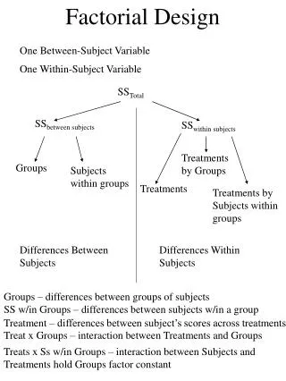
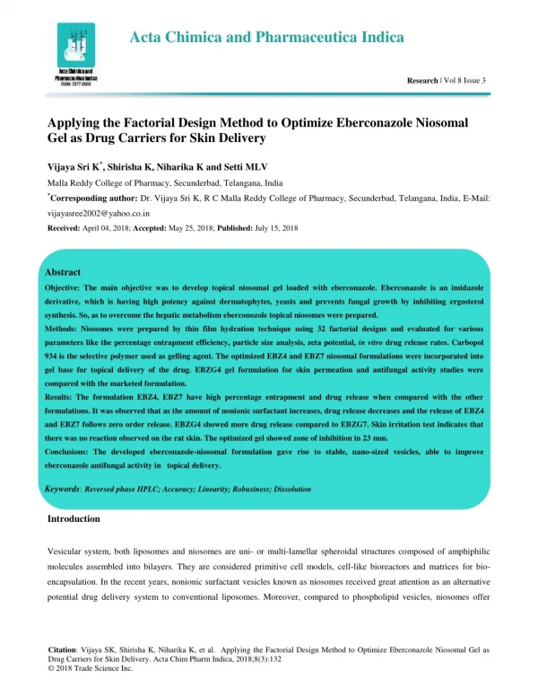
Acta Chimica and Pharmaceutica Indica Research | Vol 8 Issue 3 Applying the Factorial Design Method to Optimize Eberconazole Niosomal Gel as Drug Carriers for Skin Delivery Vijaya Sri K*, Shirisha K, Niharika K and Setti MLV Malla Reddy College of Pharmacy, Secunderbad, Telangana, India *Corresponding author: Dr. Vijaya Sri K, R C Malla Reddy College of Pharmacy, Secunderbad, Telangana, India, E-Mail: vijayasree2002@yahoo.co.in Received: April 04, 2018; Accepted: May 25, 2018; Published: July 15, 2018 Abstract Objective: The main objective was to develop topical niosomal gel loaded with eberconazole. Eberconazole is an imidazole derivative, which is having high potency against dermatophytes, yeasts and prevents fungal growth by inhibiting ergosterol synthesis. So, as to overcome the hepatic metabolism eberconozole topical niosomes were prepared. Methods: Niosomes were prepared by thin film hydration technique using 32 factorial designs and evaluated for various parameters like the percentage entrapment efficiency, particle size analysis, zeta potential, in vitro drug release rates. Carbopol 934 is the selective polymer used as gelling agent. The optimized EBZ4 and EBZ7 niosomal formulations were incorporated into gel base for topical delivery of the drug. EBZG4 gel formulation for skin permeation and antifungal activity studies were compared with the marketed formulation. Results: The formulation EBZ4, EBZ7 have high percentage entrapment and drug release when compared with the other formulations. It was observed that as the amount of nonionic surfactant increases, drug release decreases and the release of EBZ4 and EBZ7 follows zero order release. EBZG4 showed more drug release compared to EBZG7. Skin irritation test indicates that there was no reaction observed on the rat skin. The optimized gel showed zone of inhibition in 23 mm. Conclusions: The developed eberconazole-niosomal formulation gave rise to stable, nano-sized vesicles, able to improve eberconazole antifungal activity in topical delivery. Keywords:Reversed phase HPLC; Accuracy; Linearity; Robustness; Dissolution Introduction Vesicular system, both liposomes and niosomes are uni- or multi-lamellar spheroidal structures composed of amphiphilic molecules assembled into bilayers. They are considered primitive cell models, cell-like bioreactors and matrices for bio- encapsulation. In the recent years, nonionic surfactant vesicles known as niosomes received great attention as an alternative potential drug delivery system to conventional liposomes. Moreover, compared to phospholipid vesicles, niosomes offer Citation: Vijaya SK, Shirisha K, Niharika K, et al. Applying the Factorial Design Method to Optimize Eberconazole Niosomal Gel as Drug Carriers for Skin Delivery. Acta Chim Pharm Indica, 2018;8(3):132 © 2018 Trade Science Inc.
www.tsijournals.com | September-2018 higher chemical and physical stability [1] with lower cost and greater availability of surfactant classes [2]. Niosomes have been reported to enhance the residence time of drugs in the stratum corneum and epidermis, while reducing the systemic absorption of the drug and improve penetration of the trapped substances across the skin. In addition, these systems have been reported to decrease side effects and give a considerable drug release [3]. They thought to improve the horny layer properties both by reducing trans epidermal water loss and by increasing smoothness via replenishing lost skin lipids. Moreover, it has been reported in several studies that compared to conventional dosage forms, vesicular formulations exhibited an enhanced cutaneous drug bioavailability [4]. Stratum corneum (SC) is a main barrier of many compounds passing through the skin. Several approaches have been developed to weaken this skin barrier. One possibility for increasing the penetration of drugs and many cosmetic chemicals is the use of vesicular systems, such as liposomes and niosomes. Several researchers have developed novel elastic vesicles in order to deeply and easily penetrate through the skin [5,6]. Eberconazole is a topical imidazole antifungal agent which inhibits sterol synthesis and is active against most species of yeast and dermatophytes and has very good local tolerability. Eberconazole is distinct from other imidazole as it shows anti- inflammatory activity, which favors its use in the management of inflamed dermatophytic infections [7]. To avoid from hepatic toxicity, it has been formulated as topical use for the treatment of dermatophytosis. It has shown high potency against dermatophytes and yeasts. Dermatophytosis is superficial skin infection caused by dermatophytes. Since the infection is limited to the superficial layers of skin, topical application of eberconazole is helpful in the treatment. The aim of present research work was to enhance the penetration of the drug and increase the residence time at the target. Niosomes were prepared by thin film hydration technique because in this method, multilamellar vesicles are formed and entrapment efficiency is high. Niosomes with controlled release rates of drug was made into gels using carbopol 934 and the skin permeation study was conducted in comparison with the marketed cream. Further, the antifungal activity measurement was done using the eberconazole niosomal gel. Materials and Methods Materials Eberconazole gift sample from Dr. Reddy’s laboratory Hyderabad. Span 60, cholesterol and carbopol were supplied by NR chemicals limited Hyderabad. All the chemicals used were of analytical reagent (AR) grade. Preparation of eberconazole niosomes Eberconazole niosomes were prepared by thin film hydration technique following 32 factorial designs. Respective amounts of span 60 and cholesterol in different proportions were dissolved in 10 ml methanol keeping amount of drug constant. The solvent was allowed to evaporate by continuous rotating over the rotary vacuum evaporator until a thin dry film was formed along the wall of the round bottom flask, which was then hydrated with 10 ml of phosphate buffer pH 6.4 at 60ºC [8]. 2
www.tsijournals.com | September-2018 The formulation was designed to study the interaction of variables of formulation on physical characteristics of niosomes. The studied variables and the levels of each factor contain the formulation of the factorial design is represented in TABLE 1. Each row identifies an experiment and provides a result (response). The levels of the factors studied were chosen so that the irrelative difference was adequate to have a measurable effect on the response, along with the information that the selected levels are within practical use [9]. TABLE 1. Variables in 32 full factorial design Independent variable, Factor Levels used Lower (-1) Middle (0) Upper (+1) Span 60 (X1) mg 20 40 60 Cholesterol (X2) mg 20 40 60 Dependent variable, response Entrapment efficiency Amount of EBZ drug used was 10 mg in all the formulations Characterization of niosomes Microscopic examination, pH and appearance Small amounts of the formed niosomes were spread on a glass slide and examined for the vesicles structure and the presence of insoluble drug crystals using ordinary light microscope with varied magnification powers (10X and 40X). Photomicrographs were taken for niosomes using Fujifilm Finepix F40 fd 8.3 MP digital camera with 3X optical zoom. The pH of the various gel formulations was determined by using digital pH meter. All developed gels were tested for homogeneity by visual inspection. Physical parameters such as color and appearance were checked. Then gels were filled in the container and tested for their appearance and presence of any gritty particles and aggregates. Entrapment efficiency Each formulation was centrifuged at 3,000 rpm for 40 min to separate the free drug in the supernatant from the drug incorporated in the niosomes. Entrapment efficiencies were determined by complete disruption of vesicles using Triton X- 100. The drug content was determined using a spectrophotometer at 237 nm using %0.1 triton solutions as blank. Entrapment efficiency=drug entrapped/total drug*100 3
www.tsijournals.com | September-2018 Particle size, zeta potential determination and polydispersity index Vesicle properties such as particle size diameter, zeta potential and size distribution were determined by HORIBA scientific nano partica (Nano particle size analyzer) SZ-100. Zeta potential is a measure of magnitude of electrostatic or charge repulsion or attraction between particles, and is one of the fundamental parameters known to affect stability [10]. Its measurement brings detailed insight into the causes of dispersion, aggregation or flocculation. The niosomal dispersion was determined using zeta potential analyzer. The temperature was set at 25ºC. Electrophoretic mobility and mean zeta potential values were obtained directly from the measurement. The polydispersity index (PDI), which is the width of the particle size distribution curve, was determined as a measure of the homogeneity [11]. The Polydispersity of eberconazole loaded niosomes of non-uniform size was calculated from the formula, Polydispersity=[D0.9-D0.1] ÷ D0.5 Where D0.9, D0.1 and D0.5 are particle diameters determined at 90th, 50th and 10th percentile of undesired particles respectively. Assessment of eberconazole release rates from niosomes In vitro drug release studies for eberconazole niosomal solution were carried out using dialysis membrane employing two sides open ended cylinder [12]. Cellophane membrane previously soaked overnight was mounted onto one end of the cylinder; weighed amount of niosomal solution was placed uniformly on the dialysis membrane. The two sides open ended cylinder was placed in a beaker containing 50 ml of phosphate buffer saline pH 7.4. Sample (5 ml) was withdrawn by taking into consideration. The dose of the drug at predetermined intervals (0, 1, 2, 3, 4,5, 6, 7, 8, 16, 18, 20, 22 and 24 h) are replaced with fresh medium. The volume of aliquots withdrawn shows desired sink conditions with minimum dilution in the receptor compartment. The appropriate correction factor for the same is used to calculate the amount of drug released. Preparation of eberconozole niosomal gels Required quantity of carbopol 934 (1% w/w) was weighed and dispersed in small quantity of distilled water to prepare an aqueous dispersion. The dispersion was allowed to hydrate for 3-4 h. Other ingredients like propylene glycol (10% w/w) and glycerol (30% w/w) were added subsequently to the aqueous dispersion with continuous stirring. The niosomal pellet obtained after centrifugation was added to the dispersion. The dispersion was neutralized with 1% w/v sodium hydroxide solution and pH was adjusted to 6. The entrapped air bubbles were removed using vacuum and leaving the gels overnight [13]. 4
www.tsijournals.com | September-2018 Viscosity analysis The viscosity of gel was measured by using a programmable viscometer (model DV-II + Pro, Brookfield Engineering Laboratories, Inc., USA). T-bar spindle (spindle-C, S-96) was lowered perpendicularly into the gel placed in a beaker taking care that the spindle did not touch the bottom of the beaker. The spindle was rotated at a speed of 50 rpm and the readings were recorded after 30 s when the gel level was stabilized. Ex-vivo skin permeation studies Niosomes are composed of nonionic surfactants, which are biocompatible and relatively nontoxic and themselves serves as excellent penetration enhancers [14]. In this study, in order to assess the influence of the drug carriers on the accumulation into and diffusion of drug through the skin, skin permeation studies using pig ear skin. Superficial skin was taken from the back of pig ear and using a depilatory preparation hair was removed. The cleared area was washed with pH 7.4 phosphate buffer. The collected and prepared skins were tied mounted onto one end of the cylinder; weighed amount of niosomal gel and the marketed eberconazole cream were placed uniformly on the skin and the study was conducted. The two sides open ended cylinder was placed in a beaker containing 50ml of phosphate buffer saline pH 7.4. Sample (5 ml) was withdrawn at predetermined intervals and replaced with fresh medium. The volume of aliquots withdrawn presents desired sink conditions with minimum dilution in the receptor compartment and the appropriate correction factor for the same is used to calculate the amount of drug released. Antifungal activity zone of inhibition by cup plate method Sabouraud dextrose agar can be used for cultivating yeasts, moulds and acid uric microorganisms. This medium is also used for determining the microbial and fungal content of cosmetics and for the mycological evaluation of food. The formula is based on European pharmacopoeia. Dextrose is a fermentable carbohydrate providing carbon and energy. Peptone mixtures provide nitrogen, vitamins, minerals and amino acids essential for growth. The high dextrose content and acidic pH makes this medium selective for fungi. The selected Candida albicans is transferred into the prepared Sabouraud dextrose agar and incubated at 37ºc for 24 to 48 h. The antifungal activity is studied by zone of inhibition of cup plate method. For the study of zone of inhibition firstly agar is prepared. Prepared agar is transferred into petri plate and left for solidification aseptically by using laminar air flow after solidification of that agar spread the fungal solution of Candida albicans. After the whole spreading of fungal solution, prepare holes in middle by using cork borer and then the eberconazole loaded niosomal gel (EBZG4), marketed formulation, are poured into that holes by using pipette, during pouring make sure that the 5
www.tsijournals.com | September-2018 solution doesn’t get flooded. Then these Petri-plates are incubated at 37ºc for 24 to72 h, and then count the zone of inhibition [15]. Results and Discussion Microscopic examination, pH and appearance The niosomes were prepared by thin film hydration method. The prepared niosomes were discrete, (FIG. 1) nearly spherical nanometric particles in the size range of 230-470 nm. The pH values of the formulations EBZG4 and EBZG7 and marketed gel were found to be 7.2 pH. The formulated gels showed good homogeneity and no lumps in the formulation. The formulated preparations were much clear and translucent. FIG. 1. Photomicroscopic of Eberconazole niosomal solution. Entrapment efficiency Effect of cholesterol: The entrapment efficiency is the most important parameter from pharmaceutical viewpoint in niosomal formulations. Various techniques may be used to optimize the drug loading and this is very important in industrial settings. Incorporation of cholesterol was known to influence vesicle stability and permeability. It is also one of the common and essential additives in niosome formulation in the present study. 6
www.tsijournals.com | September-2018 A high percentage of entrapment would mean less time and effort involved in removal of unentrapped material. To study the effect of cholesterol on the amount of drug entrapment in niosomes, a series of formulations were prepared with different cholesterol amounts at a fixed amount of eberconazole (10 mg). The effect of cholesterol on eberconazole entrapment was varied according to the nonionic surfactant. As the HLB of the surfactant increases, the minimum amount of cholesterol necessary to form vesicles increases [10]. Niosome formulation EBZ7 prepared using Span 60 showed the maximum entrapment efficiency. Increasing cholesterol content from 20 to 60 mg lead to a significant decrease in the entrapment efficiency of eberconazole niosomes are shown in Quadratic 3d surface plot and Contour plots obtained by fixing the XC factor at its cholesterol and span and varying (XA) and (XB) over the range used in the factorial study. FIG. 2 and 3 depicts Quadratic 3d surface plot and contour plots which show the effects of XA and XB on EE%. FIG. 2. Quadratic 3d surface plot showing the effect of span and cholesterol on entrapment efficiency. FIG. 3. Contour plot for entrapment efficiency of eberconazole niosomal solution. 7
www.tsijournals.com | September-2018 The major reduction in drug entrapment when cholesterol content was further increased may be due to two conflicting factors: (1) with increased cholesterol, the bilayer hydrophobicity and stability increased [16] and permeability decreased [17] which may lead to efficiently trapping the hydrophobic drug into bilayers as vesicles formed. (2) In contrast, higher amounts of cholesterol may compete with the drug for packing space within the bilayer, hence excluding the drug as the amphiphiles assembling into the vesicles. Another study suggested that the decreasing the entrapment efficiency with increasing cholesterol ratio above a certain limit may be due to the fact that increasing cholesterol beyond a certain concentration can disrupt the regular linear structure vesicular membranes. Effect of non-ionic surfactants The HLB value of span 60 is 4.7.since the HLB value of span 60 is less cholesterol is added to make vesicles more stable. Cholesterol lends greater stability to the bilayer membrane by raising the gel liquid transition temperature of vesicles. The liquid transition temperature of nonionic surfactants (span 20 to span 60) increases from 46º to 56ºC as the hydrocarbon length is increased from C9-C15. When the gel transition temperature increases it leads to greater stability of the bilayer. Thus increased stability decreases leakage of the vesicles and stabilizes against osmotic gradients [18-20]. The entrapment efficiency was in a range of 69 to 78%. The EBZ7 formulation having high entrapment efficiency. It was observed that the percentage entrapment efficiency was significantly affected by the applied processing variables such as concentration of span 60 as well as cholesterol. Cholesterol improves the fluidity of the bilayer membrane and improves the stability of bilayer membrane in the presence of biological fluids such as blood/plasma. Span 60 having high phase transition temperature (gel to liquid transformation) and having critical packing parameter (CPP) ranging from 0.5 to 1 entrap drug molecule without any cholesterol. It was clearly indicated that with increase in concentration of span 60 percentage, entrapment efficiency was increased whereas concentration of cholesterol was inversely related to the percentage entrapment efficiency. Particle size, zeta potential determination and poly dispersity index The formulation produced particles of size less than 1000 nm, and hence characterized as nanoparticles. The zeta potential is an important parameter upon consideration of stability of the nanoparticles invitro. The value of zeta potential of the EBZ 7 (optimized drug loaded) niosomal formulation was -25.7 mV. The values of zeta potential showed prepared niosome have sufficient charge to inhibit aggregation of vesicles due to electric repulsion [21]. Presence of charge tends to increase the interlamellar distance between successive bilayers in multilamellar vesicle structure and leads to greater overall entrapped volume. The polydispersity index (PDI), which is the width of the particle size distribution curve, was determined as a measure of the homogeneity. Polydispersity index was very low it reflects uniform size particles, whereas remaining formulation has somewhat non-uniform. The PDI values of the niosomal formulations are shown in TABLE 2. 8
www.tsijournals.com | September-2018 TABLE 2.Variables in 32 full factorial design Code of formulation Span 60 Concentration (X1) -1 (20 mg) Cholesterol Concentration (X2) -1 (20 mg) EBZ1 EBZ2 -1 (20 mg) 0 (40 mg) EBZ3 -1 (20 mg) +1 (60 mg) EBZ4 0 (40 mg) -1 (20 mg) EBZ5 0 (40 mg) 0 (40 mg) EBZ6 0 (40 mg) +1 (60 mg) EBZ7 +1 (60 mg) -1 (20 mg) EBZ8 +1 (60 mg) 0 (40 mg) EBZ9 +1 (60 mg) +1 (60 mg) Assessment of eberconazole release rates from niosomes In-vitro drug release of EBZ loaded niosomes prepared using span 60 and cholesterol in various molar ratios are shown in FIG. 3. it has been observed that the niosomes prepared using span 60 and cholesterol in the molar ratios 1:1 (EBZ5) formulation showed drug release of 87.52% while the drug release of 85.33%, for the formulation EBZ7. From the results it is obvious that lipophilicity of the surfactants may determine the rate of drug release. Span 60 being relatively lipophilic impedes the easy permeation to the aqueous phase and could account for higher drug release. Skin permeation study The ex vivo diffusion data indicates that the release is diminished to a great extent as compared to the in vitro release of niosomes (EBZG4). This could be because of accumulation of drug in the skin layers. The delayed drug release rate may be attributed largely to the drug transport by diffusion controlled mechanism resulting in prolonged drug release profile. The release profile of the developed formulation data is shown in FIG. 4. The fluxes after 24 h of the formulations (EBZG4 and marketed) investigated were 30.83 and 27.35 µg/cm2h respectively. This result has supported that EBZG4 is efficiently releasing drug from the niosomal gel when compared to marketed formulation (FIG. 5). 9
www.tsijournals.com | September-2018 100 90 80 70 % Drug release 60 50 40 30 20 10 0 0 4 8 12 16 20 24 Time(hr) EBZ1 EBZ6 EBZ2 EBZ7 EBZ3 EBZ8 EBZ4 EBZ9 EBZ5 FIG. 4. Comparison of influence of formulation variables on invitro drug release of eberconazole loaded niosomes. 3500 3000 2500 CDR(µg/cm2) 2000 EBZG4 1500 MARKTD 1000 500 0 0 4 8 12 16 20 24 TIME(HRS) FIG. 5. Percentage release of optimized eberconazole niosomal gel and marketed gel Across excised pig ear skin. 10
www.tsijournals.com | September-2018 Antifungal activity measurement The niosomal gel with good entrapment and controlled release was selected for antifungal measurement and compared with the marketed product. The tests showed that the pure drug has maximum zone of inhibition of 30 mm whereas the niosomal formulation showed less zone of inhibition than pure drug. The marketed formulation and niosomal gel is having zone of inhibition of 21 mm and 23 mm. The gradual increase in zone of inhibition during the study is period due to the controlled release of medicament. The results were shown in TABLE 3. TABLE 3. Antifungal activities of eberconazole Zone of inhibition (in mm)* ± S.D Samples 24 h 48 h 72 h Pure drug 19.2 ± 0.4 23.2 ± 0.7 30.7 ± 0.9 Marketed cream 15.6 ± 1.5 19.6 ± 1.5 21.5 ± 1.1 Eberconazole loaded niosomal gel (EBZG4) 16.1 ± 1.0 20.3 ± 0.5 23.2 ± 1.1 Conclusion Controlled release niosomal gel dosage form of eberconazole was successfully developed using factorial statistical design. Dissolution studies for 24 h showed extended drug release for prolonged duration. The optimized formulation, when compared to the marketed formulation seems to be promising for improving penetration and antifungal activity of eberconazole. REFERENCES 1.Vora B, Khopade A, Jain NKJ. Proniosome based transdermal delivery of levonorgestrel for effective contraception. J Control Release. 1998;54:149-65. 2.Manconi M, Sinico C, Valenti D, et al. Niosomes as carriers for tretinoin III A study into the in vitro cutaneous delivery of vesicle-incorporated tretinoin. Int J Pharm. 2006;311:11-19. 3.Schreier H, Bouwstra J. Liposomes and niosomes as topical drug carriers: dermal and transdermal drug delivery. J Con Rel. 1994;30:1-15. 4.Mura S, Pirot F, Manconi M, et al. Liposomes and niosomes as potential carriers for dermal delivery of minoxidil. J Drug Target. 2007;15:101-08. 5.Dayan N, Touitou E. Carriers for skin delivery of trihexyphenidyl HCl: ethosomes vs. liposomes. Biomat. 2000;21:1879-85. 11
www.tsijournals.com | September-2018 6.Koskela R, Kirjavainen M, Monkkonen J, et al. Enhancement of percutaneous absorption of naproxen by phospholipids. Int J Pharm. 1998;175:225-30. 7.Subramanya L, Bangera M, Martis J, et al. Eberconazole- Pharmacological and clinical review. Ind J Derm. Venereal Leprol. 2012;78:217-22. 8.Ismail Md, Mouzam D, Shaik S. Development and characterization of Salmeterol Xinafoate niosomes for nasal delivery. Ind J Pharm Edu Res. 2011;45. 9.Mahesh N, Padamwar, Varsha B. Pokharkar. Development of vitamin loaded topical liposomal formulation using factorial design approach: Drug deposition and stability. Int J Pharm. 2006;320:37-44. 10.Devaraj GN, Parakh SR, Devraj R, et al. Release studies on niosomes containing fatty alcohols as bilayer stabilizers instead of cholesterol. J Colloid Interface Sci. 2002;251:360-65. 11.Uchegbu IF, Vyas SP. Nonionic surfactant based vesicles in drug delivery. Int J Pharm. 1998;172:33-70. 12.Vijayawagh D, Deshmukh O. Itraconazole niosomes drug delivery system and its antimycotic activity against Candida albicans. ISRN Pharm 2012;7. 13.Aliasgar S, Ambikanandan M. Studies in topical application of niosomally entrapped nimesulide; J. Pharm Pharm Sci, 2002;5:220-25. 14.Jayaprakash S. Preparation and evaluation of celecoxib transdermal patches. J Pharm Sci. 2010;23(3):279-283. 15.Wankhade S. Isolation of pure culture of bacteria from soil and study their antimicrobial activity. Pharmacolog. 2011;2:679-84. 16.Bernsdorff C,Wolff A, Winter R, et al. Effect of hydrostatic pressure on water penetration and rotational dynamics in phospholipids–cholesterol bilayers. Biophys J. 1997;72:1264-77. 17.Kirby C, Clarke J, Gregoriadis G. Effect of the cholesterol content of small unilamellar liposomes on their stability in vivo and in vitro. Biocheme J. 1980;186:591-98. 18.Joseph G. In Vitro Activities of the New Antifungal Drug Eberconazole and Three Other Topical Agents against 200 Strains of Dermatophytes. 2003;41(11):5209-11. 19.Muzzalupo R, Pérez L, Pinazo A, et al. Pharmaceutical versatility of cationic niosomes derived from amino acid- based surfactants: Skin penetration behavior and controlled drug release. Int J Pharm. 2017;529(1-2):245-52. 20.Ahmad MZ, Mohammaed AA, Ibrahim MM. Technology overview and drug delivery application of proniosome. Pharma Dev Technol. 2017:302-11. 21.Barbanoj MJ, Antonijoan R, García-Gea C, et al. Eberconazole cream: topical and general tolerability, sensitization potential, and systemic availability. Methods Find Exp Clin Pharmacol. 2005;27:227-34 12