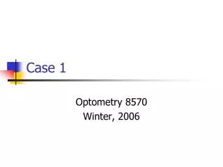
Case 1
Case 1. Optometry 8570 Winter, 2006. Mr. FM. 72 yo WM CC: “irritation and pain” HPI: OD; “couple of days” Severity varies throughout day Other symptoms include include morning discharge OU and photophobia Has been using artificial tears. More about FM.

Case 1
E N D
Presentation Transcript
Case 1 Optometry 8570 Winter, 2006
Mr. FM • 72 yo WM • CC: “irritation and pain” • HPI: • OD; “couple of days” • Severity varies throughout day • Other symptoms include include morning discharge OU and photophobia • Has been using artificial tears
More about FM • MHx: NIDDM for 10+ years, arthritis • Meds: Digoxin, Metformin, Metoprolol, Naproxen, Hydroxychlor, Methotrexate, Folic Acid, and Aspirin • Wears a near correction only • No other OHx or FOHx • LEE: 2 years ago
9-22-05 • Dist VA sc: OD 20/30-, OS 20/30-, OU 20/25 • Conf VF: full OD/OS • EOMs: FROM OU • Pupils: PERRLA, -APD 4-2 OU • IOPs: 10/10
9-22 SLEx and Fundus • OS: WNL • OD: • Gr 2 conj. Injection w/ chemosis • 3X5 mm area of infiltration with heaped epithelium and neovascularization inferior nasal with positive staining • Fundus WNL OU; C/D 0.2/0.2 OU
Questions • What’s your DDx to explain this patients signs and symptoms? • What other documentation or recordkeeping might help you with the Dx? • What type of discharge do you think is present, based on the other signs and symptoms? • What does neovascularization have to do with the Dx in this case?
The Intern Dx’ed this… • Corneal ulcer secondary to bacterial infection • Tx: • Vigamox q2h, then QID • Voltaren QID • F/U 2 days • Do you agree? Why?
2 days later… • No improvement of Sxs • Little corneal improvement • Tx: • Vigamox QID • Voltaren QID • F/U 2 days
2 days later… • Pt reports improvement in Sxs but still a little scratchy • SLEx: improvement but still some staining • Tx: • Continue Vigamox QID • D/C Voltaren • F/U 2 days • Similar findings 2 days later; no changes in Tx
1 week f/u since last visit • Pt reports eye feels much better • SLEx: still some heaped epithelium w/ staining • Pt reports he has not been compliant w/ his meds • Tx: • Restart Vigamox QID • Add Lotemax TID
2 days later • Pt reports no longer has Sxs • SLEx: Cornea much flatter w/ little to no staining • Tx: • Continue Vigamox and Lotemax • RTC 1 week • (This is the last time I saw this pt but he reportedly was still doing well and his meds were d/c)
Questions • Why was the antibiotic not effective in relieving symptoms initially? • Do you think the Dx was infectious keratitis, based on the signs, symptoms, and response to high-dose antibiosis? • What other diagnosis may explain this case, in light of the patient’s age, local associated neo, and the immediate effectiveness of the added steroid?
Student Dx next page… • (Dr. Crane does not entirely agree…)
What can cause infectious keratitis? • Bacteria is the most common cause • Neisseria gonorrhoeae and H. influenza are especially dangerous because they have the ability to penetrate intact epithelium • Other species include: Staphylococcus, Streptococcus, Pseudomonas, and Moraxella • Cornea may be compromised by contact lens wear, pre-existing corneal diseases, chronic blepharoconjunctivitis, chronic dacryocystitis, tear film deficiency, and topical steroid therapy
Signs and Sxs • Sxs: foreign body sensation, photophobia, blurred vision, pain, eyelid edema, discharge, red eye, and contact lens intolerance • Signs: conjunctival and circumcorneal injection, epithelial or stromal infiltrate, mucopurulent discharge, stromal edema, anterior chamber reaction with or without hypopyon, corneal thinning, folds in Descemet’s membrane, and eyelid edema
Differential Dxs • Differential keratitis diagnoses may be fungal, acanthamoeba, HSV, atypical mycobacteria, and sterile corneal infiltrates • Fungal keratitis is characterized by feathery borders • Acanthamoeba is generally extremely painful; it is usually associated with contact lens overwear or swimming in contact lenses • HSV is often recurrent with epithelial dendrites • If the infection follows injury or surgery it may be caused by atypical mycobacteria
DDx • Dr. Crane’s proposed Dx does not appear on the list on the last slide. • What Dx do you think is missing?
The Exam • Complete history- including contact lens history, trauma, previous eye care, eye disease, and systemic illnesses • SLEx- pay particular attention to the cornea with and without stain, anterior chamber reaction, and IOP measurement • Culture- Large infiltrates, central infiltrates, infections which are unresponsive to treatment
Tx • Topical antibiotics are the primary form of treatment • A broad-spectrum topical antibiotic such as Moxifloxacin, Gatifloxacin, or Ciprofloxacin should be the medication of choice • At first, the medication should be delivered every hour to every two or four hours • Cycloplegic drops may be used for comfort, to prevent synechiae formation, or prevent hypopyon formation • Topical analgesic may also be used for discomfort • Topical corticosteroid may be used only once the infection is under control and there is noticeable improvement • F/U daily at first, then every couple days, then weekly • There should be particular attention paid to size and depth, symptoms, and the anterior chamber reaction
Conclusions about this case • FM presented to the office with primary complaints of pain, irritation, and light sensitivity. Bacterial Keratitis can cause a patient many problems if not treated quickly and correctly. This patient was difficult to treat. The cornea did not appear to respond quickly to the treatment with Vigamox and Voltaren. However, since the patient is a diabetic, a slower healing time was expected. Once the epithelium began to heal, the patient was put on Lotemax. This seemed to speed up the healing process. It is very important to get these infections under control before it begins to affect the visual axis.
Do you agree with the intern’s statements above? • Is the fact that he’s diabetic the only reason why Vigamox did not initially work? • What is your final Dx?
References • Kunimoto, Kantikar, and Makar. The Wills Eye Manual. Philadelphia, 2004: pages 52-56. • Kanski, Jack. Clinical Ophthalmology. Washington, 2003: pages 102-105.