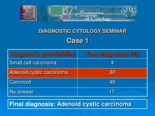
Case 1
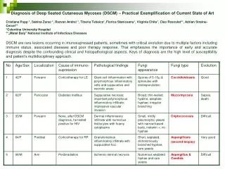
Diagnosis of Deep Seated Cutaneous Mycoses (DSCM) – Practical Exemplification of Current State of Art Cristiana Popp *, Sabina Zurac *, Razvan Andrei *, Tiberiu Tebeica *, Florica Staniceanu *, Virginia Chitu *, Cleo Rosculet **, Adrian Streinu-Cercel **

Case 1
E N D
Presentation Transcript
Diagnosis of Deep Seated Cutaneous Mycoses (DSCM) – Practical Exemplification of Current State of Art Cristiana Popp *, Sabina Zurac *, Razvan Andrei *, TiberiuTebeica*, FloricaStaniceanu*, Virginia Chitu*, Cleo Rosculet**, Adrian Streinu-Cercel** *Colentina University Hospital **„MateiBalș” National Institute of Infectious Diseases DSCMare rare lesions occurring in imunosupressed patients, sometimes with critical evolution due to multiple factors including immune status, associated diseases and poor therapy response. That emphasizes the importance of early and accurate diagnosis despite the confounding clinical and histopathological aspects. Keys of diagnosis are the high level of susceptibility and patient’s multidisciplinary approach.
Case 1 HE PAS Case 2 HE Grochott The fungal elements are rather inconspicuous in HE stain. Very important for diagnosis is the routine examination of at least one fungal stain for each inflammatory cutaneous lesion in immunosupressed patients (PAS, Grochott)
HE HE HE HE PAS Grochott Grochott Grochott Grochott PAS Case 4 Case 5 Case 3
Protocols of British Society for Medical Mycology • The pathologist is not called to establish the exact type of fungi, the pathological report must include: • the presence and absence of yeast forms, • the presence and absence of hyphae, • whether hyphae are septate or aseptate, • presence of melanin, • the size of fungi, • cellular location • any specialised structures The appearance of fungi can be sufficient to guide treatment, but the golden standard requires either immunohistochemical confirmation of the specific type or confirmatory cultures. Patients with poor immune status have, usually, a long, difficult evolution, with possible fatal outcome due to systemic dissemination of fungal infection. HE Bronchopneumonia (case 2) Applying these standards in routine examination of inflammatory skin biopsies micotic infection can be identified as cause of inflammation, thus improving management of immunocompromised patients.