Case 1
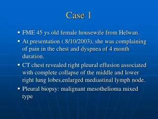
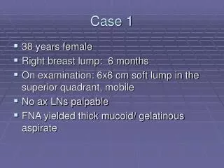
Case 1. DP 6 year old male well until morning of admission on July 29 th developed fatigue, abdominal pain, lethargy , increased sleepiness and work of breathing No fever, vomiting or diarrhea, no history of prodromal illness noted

Case 1
E N D
Presentation Transcript
Case 1 DP 6 year old male well until morning of admission on July 29th developed fatigue, abdominal pain, lethargy , increased sleepiness and work of breathing No fever, vomiting or diarrhea, no history of prodromal illness noted Upon arrival to ED, physical exam remarkable for evidence of circulatory collapse and respiratory arrest and patient intubated CV exam- normal S1, single S2 no thrill no murmur, impaired perfusion in lower extremities Respiratory exam- clear , no rales Abdominal exam - soft , non tender Liver 1 cm below RCM Due to evidence of circulatory collapse ,patient was transferred to LCH
Echocardiogram markedly dilated heart with ejection fraction of 15 %, mural thrombus was present Echocardiogram
Diagnostic Evaluation EKG Tachycardia, prolonged QT interval ,intermittent ectopic atrial foci Cardiac biopsy Minimal lymphocytes, no evidence of necrosis Laboratory evaluation CMV, EBV, Viral DFA all negative
Hospital Management Ionotropic Support Milrinone Immunosuppressive Therapy IVIG Anticoagulant therapy Heparin Adjunct therapy ECMO Discharged on Hospital Day # 26 Aspirin Enanapril Lasix Echocardiogram on discharge EF 50%
Follow Up Office visit 9/12/ 2011 – doing well on all meds Echocardiogram - EF 44% calculated Office visit 12/15/2011 – history of tachycardia at home Echocardiogram - EF 27% EKG - SVT with atrial ectopic rhythm Admission 12/16/2011 Cryoablation of ectopic atrial foci, Echocardiogram EF post procedure 37% Office visit 1/13/ 2012 – history of normal heart rate EKG - normal sinus rhythm Echocardiogram EF 50%
Case 2 MG 14 year old boy with a prodromal viral illness and two week history of episodic chest pain without radiation, no fatigue, nausea ,vomiting or abdominal pain. Activity on day of admission was snowboarding without any difficulty Pt had witnessed cardiac arrest on ski slopes of Asheville CPR performed , AED delivered 5 shocks, patient intubated, placed on ionotropic medications and transferred to LCH CV exam- normal S1, S2 no thrills , rubs gallops or murmur Abdominal exam - soft non tender, no palpable liver edge Respiratory exam- no rales
Diagnostic Evaluation Echocardiogram EF 25% EKG Borderline sinus tachycardia Cardiac Enzymes Troponin 8.9 Viral Cultures CMV, EBV, viral DFA negative
Hospital Management Ionotropic Support Dopamine Epinephrine Amidarone Immunosuppressive Therapy IVIG given in Asheville
Follow Up Transfer on Hospital Day # 8 Echocardiogram EF improved to 45% Neurologic evaluation MRI - Anoxic brain injury EEG - Encephalopathy
Case 3 HN 6 day old infant admitted for fever and loose stools, admitted to hospital with entire sepsis workup done including CXR. Pt remained febrile in spite of antibiotic therapy for three days and then developed respiratory distress and tachycardia. CV exam- normal S1, single S2 no thrill no murmur, Respiratory exam- clear , no rales with mild retractions Abdominal exam - soft , non tender no hepatosplenomegaly
Diagnostic Evaluation EKG Tachycardia, ST changes and Q waves Echocardiogram Biventricular dysfunction EF 35% Laboratory evaluation CBC – lymphocytic predominance CMV, EBV, Viral DFA all negative Coxsackie B4- isolated from stool
Hospital Course Ionotropic Support Milrinone Dopamine Amiodarone Immunosuppressive Therapy IVIG Adjunct therapy ECMO Left Ventricular Assist Device Expired on Hospital Day # 32
Case 4 JM 31/2 month old with history of fever, poor feeding, vomiting and diarrhea andrespiratory distressfor 3 days CV exam- normal S1 single S2, no murmur a gallop was present peripheral pulses were decreased Resp exam- bibasilar rales Abdomen-soft ,non tender Liver 5cm below RCM
Echocardiogram Echocardiogram markedly dilated with severely deceased shortening with an EF 26%
Diagnostic Evaluation/Hospital Management EKG Sinus tachycardia with low voltage Laboratory evaluation CMV, EBV, Viral DFA all negative Ionotropic Support Milrinone Lasix
Follow Up Discharged on Hospital Day #20 ASA Lasix Spirolactone Enalapril Cardvediol Echocardiograms EF 15-20% June 1st EF 15% June 30th Transferred care to PCP and cardiologist in New Jersey – patient to be listed on transplant list
Case 5 AP 2.5 year old with known history of seizure disorder and infantile spasms on a ketogenic diet and Klonopin with three day history of emesis ,cough ,low grade temp seen by PCP thought to have viral process and given supportive care recommendations and follow appointment next day. Overnight, pt developed respiratory distress and went to ED On evaluation in ED , patient was noted to have respiratory failure and cardiovascular collapse was subsequently intubated and transferred to LCH CV exam S1 single S2 , S4 at LSB and cool extremities Abdomen – soft non tender Liver 2 cm below RCM Respiratory exam - clear chest no rales with chest tube in place in Left side
Diagnostic Evaluation Echocardiogram Marked dilation, decreased function on left EKG Low voltage Tachycardia, non specific T wave changes Laboratory evaluation Rapid Influenza positive CMV, EBV, Viral DFA all negative Tracheal aspirate positive for Pseudomonas
Hospital Management Ionotropic Support Milrinone Dopamine Lasix Arrhythmia management Amiodarone Immunosuppressive Therapy IVIG
Hospital Management Discharge on Hospital Day # 14 Lasix Spirolactone Enalapril Echocardiogram EF 35%
Follow Up Office visit 4/13/2011 – had run out of meds off meds for 2 weeks without signs of compromise Echocardiogram- EF 50% Office visit 6/06/2011 – off all cardiac meds Echocardiogram – EF 62%
Pediatric Myocarditis Myocarditis in Children • Defined as inflammation of the myocardium resulting in necrosis and/or degeneration of the myocyte • Inflammation may be diffuse or local • Infectious etiology more common in children, with viral infections being more common in US. • Wide variation of clinical features can complicate recognition • Most common cause of heart failure in otherwise healthy children
Pediatric Myocarditis at LCH From January 2010 to December 2011, more than 25 children admitted for myocarditis The majority of LCH cases are of suspected infectious etiology Viral isolates from LCH include Influenza A, EBV, Coxsackie B4, Parvovirus
Incidence and Prevalence • Myocarditis is rare, true incidence is unknown • Subclinical cases • Unexpected deaths • Pediatric case studies estimate a annual incidence of 1 per 100,000 children • Cases may be sporadic or epidemic with seasonal and geographical variation • Myocarditis is more common in children than adults and more often presents as acute or fulminant disease • Children also more likely to have sudden death • On post mortem examination, 12 - 17 % of sudden death without history suggestive of myocarditis showed evidence of active or healed myocardium inflammation (versus 1% to 9% of adults)
Etiologies of Myocarditis • Most commonly identified viruses • Coxackie A and B Virus • Adenovirus • Hepatits C • CMV • Echovirus • Influenza • EBV • Parvovirus B19 • HIV • Other Infectious • Bacterial, Fungal, Protozoan and Parasitic, Rickettsial • Non-Infectious Causes • Systemic Disorders • Connective tissue disorders • Cardiotoxins • Hypersensitivity reactions • Radiation
Fulminate Myocarditis Antecedent viral prodrome is variable (10 – 80 %) Acute onset of severe hemodynamic compromise, presenting as severe CHF or cardiogenic shock. Malignant arrhythmias are more common, may result in sudden death Acute Myocarditis Less distinct onset and less hemodynamic compromise Often worse outcome than fulminant with progression to failure and need for transplant or death more common Chronic Myocarditis - uncommon Clinical Manifestations
History • Infants and young children may have nonspecific signs and symptoms • Pneumonia, bronchiolitis, or asthma symptoms • Failure to thrive or feeding intolerance • Gastroenteritis symptoms with anorexia, vomiting and diarrhea • Lethargy • Inability of infants and children to communicate symptoms
History Older children/Adolescents Chest or abdominal pain was only complaint in 30% adolescents Fatigue is common Exercise intolerance, dyspnea and orthopnea also common Atypical manifestations including syncope or seizures
Physical Examination • CNS findings • Altered mental status secondary to hypoperfusion • Lethargy • Irritability • Respiratory findings • Tachypnea • Retractions • Wheezing, coughing, grunting or nasal flaring
Physical Examination • Cardiac findings • Tachycardia, esp. out of proportion for age • S3 (ventricular gallop) • Murmurs • Poor pulses and perfusion with cool extremities and decreased capillary refill • Hypotension • Distant heart sounds with pericarditis • Abdominal findings • Hepatomegaly
Diagnostic Evaluation Chest Radiograph Cardiac silhouette Enlarged in acute or chronic myocarditis Maybe normal in fulminant myocarditis Normal cardiac silhouette does not rule out myocarditis Pulmonary vascular congestion
Diagnostic Evaluation Electrocardiography Can be normal or abnormal Sinus tachycardia most common abnormality Low voltage QRS complexes, nonspecific ST and T wave changes Left ventricular hypertrophy with repolarization changes. Supraventricular/ventricular tachycardia Ventricular ectopy or heart block
Diagnostic Evaluation • Echocardiogram • Rarely normal in myocarditis • Left ventricular > right ventricular dysfunction • Decreased ejection fraction, ventricular dilatation • Mitral > Tricuspid valve regurgitation • Wall motion abnormalities • Ventricular thrombi
Diagnostic Evaluation • Endomyocaridal Biopsy (EMB) • Gold standard for diagnosis • Dallas criteria • “Active Myocarditis” includes mononuclear inflammatory cell infiltrate of myocardium and necrosis of adjacent myocytes • “Borderline myocarditis” if infiltration without myocyte destruction • 5 or more tissue specimen samples
Diagnostic Evaluation • Limitations of EMB • Inherent risk of cardiac catheterization • Limited sensitivity (20 to 50%) and specificity due to sampling error • Expert interpretation may vary • Usefulness of EMB • Unexplained cardiomyopathy • Differentiate viral myocarditis from other primary dilated cardiomyopathy (CDM) • May be used for viral PCR or viral culture
Diagnostic Evaluation Cardiac MRI Useful to document location and extent of inflammation. Has shown to increase endomyocardial biopsy sensitivity Can be used to follow progression
Laboratory Studies • Cardiac enzymes • Cardiac Troponin I or T • 35% of suspected myocarditis cases are elevated • Correlates with myocardial inflammation on EMB • CK-MB • Nonspecific inflammatory markers • ESR, CRP • Elevated WBC with lymphocyte predominance
Laboratory Studies • Viral Studies • Viral PCR • Acute and convalescent viral antibody titers for adenovirus, EBV, CMV • Stool culture for enterovirus • Tracheal aspirate viral PCR/culture
Critical Care Management • Inotropic therapy • Dopamine, dobutamine, milrinone • Used with caution as may instigate or worsen arrhythmia in unstable myocardium • Afterload reduction • Milrinone • Arrhythmia management • Adenosine, amiodarone, lidocaine
Critical Care Management • Respiratory Support • Tissue oxygenation, ventilation • Anticoagulation • Temporary transvenous or epicardial pacemaker • Conservative fluid management • Preserve preload without overload • Diuretics
Immunosuppressive Therapy • Immunosuppression • May shorten course and lessen severity, but no randomized controlled trials • Single dose IVIG 2 g/kg has shown improved left ventricular function and 1-year survival rates in EMB proven pediatric myocarditis • Steroids tapered over 2 months • Azathioprine • Cyclosporine • Muromonab (OKT3) targeting myocardial Coxsackie-Adenovirus receptor (CAR)
Adjunct Therapies • Extracorporeal Life Support (ECMO) • Indications include escalating inotropic support, ongoing metabolic acidosis and elevated serum lactate • Limited to 2 weeks duration • Ventricular assist devices • Can provide long term support with excellent quality of life • Use for smaller children/infants in US is experimental
Prognosis • Long term survival rate in children is 80% • Severity of presentation does not necessarily predict long term outcome • Highest mortality in the first 72 hours • Unfortunately long term survival does not imply full recovery • Progression to dilated cardiomyopathy (DCM) decreases survival rates to 60% at one year and 35% at 5 years
References Viral myocarditis in Children. Tammy L. Uhl. Critical Care Nurse, 2008. Vol 28: pp 42-68. Myocarditis presenting as gastritis in children. Yi’Jung Chang et al. Pediatric Emergency Care, Vol 22: No 6: pp 439-440. Immunosupressvie therapy in acute myocarditis: an 18 year systemic review. C.P. P. Hia et al. Archives of Diseases in Children. 2004. Vol 89: pp 580-584. Incidence of fatal myocarditis: A population based study in Finland. Ville, Kyto. Americal Journal of Epidemology. 2007, Vol. 165: pp 570-574. Myocarditis: Trends in Diagnosis and Treatment. Jarred Magnani and William Dec. Circulation 2006. Vol 113: pp 876-890. Diagnosis and Treatment of Viral Myocarditis. Jason Schultz et al., Mayo clinic Proceedings. Nov 2009. Vol 84: pp 1001-1009. Detection and Evaluation of Asymptomatic Myocarditis in Schoolchildren: Report of Four Cases. Clinical Investigations: Cardiology. Vol 116: August 1999; pp 340-345. Management and Outcome of Pediatric Patients Presenting with Acute Fulminant Myocarditis. Sarah Teele et al., Journal of Pediatrics. Vol 158: No 4; pp 638-645. A formidable challenge: The diagnosis and treatment of viral myocarditis in children. Derek Wheeler et al. Critical Care Clinics. Vol 19: 2003; pp 365-391