Download
1 / 31
310 likes | 327 Views
Explore the intricate immune and lymphatic system, lymphocytes attacking cancer cells, and pathways of attack against pathogens. Enhance learning with curated videos, tutorials, and interactives. Understand the body's defense mechanisms against various invaders, from viruses and bacteria to cancer cells. Dive into the three lines of defense - Innate, Adaptive, and Acquired Immunity, and the roles of leukocytes and antibodies in protection. Discover the importance of the immune system and its responses to external and internal threats. Uncover the function of key players in the blood cells and learn how leukocytes combat infections.

E N D
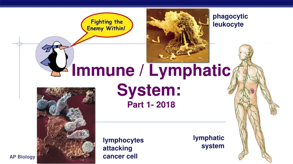
phagocytic leukocyte Fighting theEnemy Within! Immune / LymphaticSystem: Part 1- 2018 lymphatic system lymphocytes attacking cancer cell
Videos, Tutorials, Interactives • This lecture series (professor at OU) is really good. I may show at least one of her lectures 1- https://www.youtube.com/watch?v=CG931UYMbN0 2- https://www.youtube.com/watch?v=sYjtMP67vyk (this one) • 3- https://www.youtube.com/watch?v=J0Y1MlanzCk • Pretty good one from McGraw-Hill • Show this at the start of Day 2: https://www.youtube.com/watch?v=zQGOcOUBi6s • Students must complete in class (there will be questions to complete as you do the interactives): • Pearson: Ch 43 MicroFlix • HHMI- Click and Learn. • Immune system game by NobelPrize.org
Avenues of attack • Points of entry • digestive system • respiratory system • urogenital tract • break in skin • Routes of attack • circulatory system • lymph system
Why an immune system? • Attack from outside • lots of organisms want you for lunch! • animals are a tasty nutrient- & vitamin-packed meal • cells are packages of macromolecules • animals must defend themselves against invaders (pathogens) • viruses • HIV, flu, cold, measles, chicken pox • bacteria • pneumonia, meningitis, tuberculosisLyme disease • fungi • yeast (“Athlete’s foot”…) • protists • amoeba, plasmodium (malaria) • Attack from inside • cancers = abnormal body cells Mmmmm, What’s in your lunchbox?
Lines of defense • 1st line (Innate): Non-specific barriers • broad, external defense • “walls & moats” • skin & mucous membranes • 2nd line (Innate): Non-specific patrols • broad, internal defense • “patrolling soldiers” • leukocytes = phagocytic WBC • 3rd line (Adaptive/ Acquired): True immune system • specific, acquired immunity • “elite trained units” • This is also called ‘adaptive immunity’ (vertebrates) • lymphocytes & antibodies • B cells & T cells
Figure 43.2 Recognition of traits sharedby broad ranges ofpathogens, using a smallset of receptors Recognition of traits specific to particularpathogens, using a vastarray of receptors • • Bacteria & insectsinherit resistance. Vertebratesacquire immunity. Pathogens(such as bacteria,fungi, and viruses) Barrier defenses: INNATE IMMUNITY(all animals) SkinMucous membranesSecretions *NKC is analogous to Cytotoxic T cell Internal defenses: Phagocytic cellsNatural killer cellsAntimicrobial proteinsInflammatory response • Rapid response Humoral response: ADAPTIVE IMMUNITY(vertebrates only) Antibodies defend againstinfection in body fluids. Adaptive = Acquired Cell-mediated response: Cytotoxic cells defendagainst infection in body cells. • Slower response
Differentiation of Key Players (blood cells) LEUKOCYTES (WBC)
FYI: Previous Diagram • NK = Natural Killer Cells • RBC (erythrocytes)- not nucleated & have hemoglobin (carry oxygen); platelets- cell fragments (clotting) • Leukocytes means WBC, however, some books differ on the specificity. Some will use the term when referring to cells derived from myeloid progenitor myeloblast (monocytes, neutrophils, basophils, eosinophils). Others will include the lymphocytes (B & T cells). Don’t worry about this… • These WBC are phagocytic cells: monocytes, macrophages, neutrophils, dendritic cells (*need to know) • Monocytes and macrophages are basically the same thing (won’t have to differentiate for this course). FYI: monocytes are only in the bloodstream; macrophages are in other tissue • 60-70% of WBC are neutrophils. They’re usually ‘quick’ to arrive, but have short ‘lifespan’ • Basophils- inflammatory response • Eosinophils- fight parasites (like worms)
Figure 43.7 Interstitialfluid Bloodcapillary Adenoid Tonsils LYMPHATIC SYSTEM Lymphaticvessels Thymus Lymphatic vessel Tissuecells Lymphatic vessel Peyer’spatches(smallintestine) Spleen Lymphnodes Appendix(cecum) Lymphnode Masses ofdefensive cells
Innate Immunity • Video: • https://www.youtube.com/watch?v=08_wFZYj6Ww • Lecture: • https://www.youtube.com/watch?v=93J3__WC3us
1st line: Non-specific External Defense Lining of trachea: ciliated cells & mucus secreting cells • Barrier • skin • Traps • mucous (& membranes), cilia,hair, ‘earwax’ • Elimination • coughing, sneezing, urination, diarrhea • Unfavorable pH • stomach acid, sweat, saliva, urine • Lysozyme enzyme • digests bacterial cell walls • tears, sweat
2nd line: Non-specific patrolling cells bacteria • Patrolling cells & proteins • attack pathogens, but don’t “remember” for next time. Fast response • leukocytes • phagocytic white blood cells • Watch: https://www.youtube.com/watch?v=CEOV-SFTlpY • macrophages, neutrophils, natural killer cells • complement system • Free-floating proteins that destroy cells • inflammatory response • increase in body temp. • increase capillary permeability • attract phagocytes macrophage yeast
Leukocytes Involved in 2nd Line of Defense • Attracted by chemical signals released by damaged cells • Neutrophils • most abundant WBC (~70%) • ~ 3 day lifespan • phagocyte • Macrophages • “big eater” phagocyte • longer-lived • Natural Killer Cells • destroy virus-infected cells & cancer cells. • *NOTE- NKC are derived from lymphoid cells
Destroying cells gone bad! • Natural Killer Cells perforate cells • release perforin protein • insert into membrane of target cell • forms pore allowing fluid to flow in & out of cell • cell ruptures (lysis) • Watch: https://www.youtube.com/watch?v=HNP1EAYLhOs vesicle natural killer cell perforin cell membrane perforinpuncturescell membrane cell membrane virus-infected cell
Anti-microbial proteins • Complement system • ~20-30 proteins circulating in blood plasma • attack bacterial & fungal cells • form a membrane attack complex • perforate target cell • cell lysis extracellular fluid complement proteinsform cellular lesion plasma membrane of invading microbe complement proteins bacterial cell
Interferons • Proteins produced by viral infected cells • Interferon proteins are sent to nearby cells and, via signal transduction & protein synthesis, they produce enzymes that are inactive. If a virus invades, they become active and hydrolyze the viral nucleic acid. This limits ‘cell to cell’ spread of viruses • Pharmaceutical companies use recombinant DNA technology to mass produce interferon to help treat viral infections, such as Hepatitis C
Inflammatory response • Damage to tissue triggers local non-specific inflammatory response • Cells in damaged tissue release chemical signals • Such as Histamines, cytokines & prostaglandins • Causes capillaries to dilate & become more permeable (leaky) • This delivers macrophages & platelets (clotting) to tissue • fight pathogens • clot formation • increases temperature • decrease bacterial growth • stimulates phagocytosis • speeds up repair of tissues Pus- pathogens & WBC
Figure 43.8-1 Pathogen Splinter *Mast cells in damaged tissue produce signaling molecules (such as histamine and cytokines) to ‘alert’ other WBC (such as neutrophils) to come quick for battle! Macro-phage Signalingmolecules Mastcell Capillary Neutrophil Redblood cells
Figure 43.8-2 Pathogen Splinter The release of histamine (signaling molecule) causes dilation of capillaries. This allows phagocytic cells to get to the tissue to engulf pathogens. It also causes blood plasma to ‘leak’ (by osmosis) into tissue (fluid is then called interstitial fluid) and this causes swelling (edema) Movementof fluid Macro-phage Signalingmolecules Mastcell Capillary Neutrophil Redblood cells Short BBC animation- phagocytes: https://www.youtube.com/watch?v=CEOV-SFTlpY
Figure 43.8-3 Pathogen Splinter Movementof fluid Macro-phage Signalingmolecules Mastcell Phagocytosis Capillary Neutrophil Redblood cells
Fever • When a local response is not enough • system-wide response to infection • activated macrophages release interleukin-1 • triggers hypothalamus in brain to readjust body thermostat to raise body temperature • higher temperature helps defense • inhibits bacterial growth • stimulates phagocytosis • speeds up repair of tissues • causes liver & spleen to store iron, reducing blood iron levels • bacteria need large amounts of iron to grow • Watch: (1st & 2nd lines of defense-Immune Rap) https://www.youtube.com/watch?v=wgFp_U3PLBw
3rd line: Acquired (Adaptive) Immunity • Specific defense with memory (slower response than innate) • lymphocytes • B cells • antibodies(a.k.a. immunoglobulins) • T cells • Cell Mediated (target cells already infected) • Responds to… • antigens • cellular name tags • specific pathogens • specific toxins • abnormal body cells (cancer) B cell
How are invaders recognized? • Antigens • cellular ‘name tag’ proteins • “self” antigens • no response from WBCs • “foreign” antigens • Elicit specific response from WBCs (unrecognized glycoprotein markers) • pathogens: viruses, bacteria, protozoa, parasitic worms, fungi, toxins • non-pathogens: cancer cells, transplanted tissue, pollen “self” “foreign”
bone marrow Lymphocytes • B cells • mature in bone marrow • humoral response system • “humors” = body fluids • attack pathogens still circulating in blood & lymph • produce antibodies • T cells • mature in thymus • cellular response system • Cell mediated • attack infected (invaded) cells • “Maturation” • learn to distinguish “self” from “non-self” antigens • if react to “self” antigens, cells are destroyed during maturation
Humoral (antibody-mediated) immune response Cell-mediated immune response Key Figure 43.20 Antigen (1st exposure) Stimulates Engulfed by Gives rise to Antigen-presenting cell Helper T cell Cytotoxic T cell B cell Memoryhelper T cells Antigen (2nd exposure) Memorycytotoxic T cells Active cytotoxic T cells Plasma cells Memory B cells Secretedantibodies Defend against extracellularpathogens Defend against intracellularpathogens and cancer
B cells • Attack, learn & remember pathogens circulating in blood & lymph • Produce specific antibodiesagainst specific antigen • Types of B cells • plasma cells • immediate production of antibodies • rapid response, short term release • memory cells • continued circulation in body • long term immunity
Y Y Y Y Y Y Y Y Y Y Y Y Y Y Y Y Y Y Y Y Y Y Y Y Y Y Y Y Y Y Y Y Y Y Y Y Y Y Y Y Y Y Y Y Y Y Y Y Y Y Y Y Y Y Y Y Y Y Y Y Y Y Y Y Y Y Y Y Y Y Y Y Y Y Y Y Y Y Y Y Y Y Y Y Y Y Y Y Y Y Y Y Y Y Y Y Y Y Y Y Y Y Y Y Y Y Y Y Y Y Y Y Y Y Y Y Y Y Y Y Y Y Y Y Y Y Y Y Y Y Y Y Y Y Y Y invader(foreign antigen) B cells + antibodies memory cells “reserves” recognition capturedinvaders Y Y Y Y Y Y Y Y Y Y Y Y Y Y Y Y Y Y macrophage Y Y Y Y Y Y Y Y Y Y clones 1000s of clone cells plasma cells release antibodies 10 to 17 days for full response B cell immune response ‘tested’ by B cells (in blood & lymph)
TOMORROW: Immune system- part 2*show thEsE at beginning of class: https://www.youtube.com/watch?v=zQGOcOUBi6s ; Bozeman: https://www.youtube.com/watch?v=z3M0vU3Dv8E