Download

1 / 16
160 likes | 440 Views
Review of K Channels in Neuronal Action Potentials. Neurons are normally more permeable to K than Na , leaving the resting membrane potential about 65mV. Synaptic or receptor potentials cause the voltage across the membrane to become more positive. If a specific threshold is reached, an acti

E N D
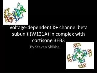
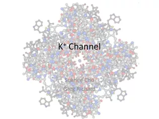
1. Crystal Structure of a Mammalian Voltage-Dependent Shaker Family K+ Channel Stephen B. Long, Ernest B. Campbell, Roderick MacKinnon
2. Review of K+ Channels in Neuronal Action Potentials Neurons are normally more permeable to K+ than Na+, leaving the resting membrane potential about
�65mV.
Synaptic or receptor potentials cause the voltage across the membrane to become more positive. If a specific threshold is reached, an action potential will occur,leaving the membrane more permeable to Na+ for a very short period. The membrane is depolarized and becomes more positive (+58mV)
3. The voltage sensors of the K+ channels sense the change to a very positive membrane potential, and they become activated.
The �inactivation peptide� that normally blocks the pore is released, and K+ ions move out of the cell in order to restore the resting membrane potential to �65mV.
The Kv1.2 channel produces a sustained delayed rectifier current,which returns the membrane potential back to it�s resting potential soon after an action potential.
These channels set the firing rate for action potentials because a consecutive one cannot be fired until the K+ channel has reset the membrane potential to it�s negative value.
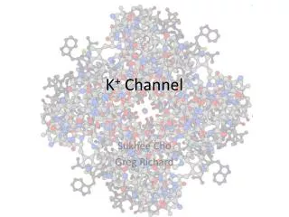
4. The Kv1.2 channel has 4 subunits that come together to form a single functional unit.
Each subunit contains 6 transmembrane helices, and 2 connector helices (S1-T1, and S4-S5)
S1-S4 helices from each subunit form the voltage sensor, and the S5 and S6 helices form the pore.
S4 -S6 helices make up the selectivity filter of the inner pore.
5. The Kv1.2 channel was co-expressed in yeast with the b2 K+ channel b subunit from a rat brain
The proteins were crystallized by vapor diffusion, and then frozen in liquid nitrogen
The resolution is 2.9A
The electron density of the b and T1 subunits was strong (B factor= 59A2), these two domains are relatively immobile compared to the pore and voltage sensor-which was very mobile(B factor=162A2).
6. Due to the weak electron density of the voltage sensor, it was constructed using prior knowledge of a KvAP (a prokaryotic channel)
The voltage sensor and pore make up the transmembrane region of the protein
The T1 and b subunits are on the intracellular side
7. The S6 inner helix on the intracellular side has a conserved Pro-Val-Pro sequence (figure B) which is essential to the gating function. This sequence is responsible for the curving of the inner helix.
The curving is responsible for coupling the voltage sensor to the pore in all Kv channels.
8. Figure 2C shows that the voltage sensor from 1 subunit is adjacent to the pore (S5and S6 of a different subunit).
The voltage sensor and the pore domain of each subunit are connected by an S4-S5 linker helix, which is parallel to the intracellular membrane at the same depth as the S6 helices that form the inner pore.
The inner pore constricts and expands, opening and closing the pore.
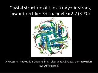
9. The Ion Conduction Pore Comparing Kv1.2 to the known structures of prokaryotic K+ channels, they concluded that the structure of the selectivity filter stays the same on the extracellular side, producing selective ion conduction.
The inner pore between the selectivity filter and the intracellular space changes it�s conformation, allowing the pore to open and close (showed by adding site directed Cis to the S6 helices.
10. Since the structures of 4 prokaryotic K+ channels are known, based on dimensions of the inner pore, the KcsA channel is closed (gray in figure 3), and the KvAP is known to be open(blue in the figure).
The pore of these K+ channels were superimposed on that of Kv1.2 (red); the Kv1.2 is deduced to be open.
This shows that there is a noticeable difference in position of the inner helices when it goes from open to closed.
11. Before the first membrane spanning helix (S1), the N-terminus forms a T1 domain inside the cell.
The T1 domains of each subunit form a tetramer at the membrane surface inside the cell.
Since the T1 domain is directly over the pore,side portals which are above the T1 domain are formed due to the T1-S1 helix linker going outward from the central axis of the channel, forming a wide space between T1 and the transmembrane domain.
12. Side portals allow for communication between the transmembrane pore and the cytoplasm , and allow K+ to flow through the channel and entry of the inactivation peptide to allow blocking of the pore.
They also allow the inner helices to move and cause opening of the pore without interference from the T1 domain.
13. The T1 domain of the channel is connected to the b-subunit, which is a tetramer of proteins
These proteins are oxido-reductases that depend on NADPH
The enzyme�s active site has a NADP+ cofactor and catalytic residues for a hydride transfer.
It is unknown whether the enzyme regulates the channel as a sensor, or if the channel regulates the enzyme
14. The function of the b-subunit is still unknown, some think that they are chaperones for the channel because they have been known to influence levels of channel expression
If the b -subunit is a �redox sensor�, and does in fact have catalytic activity, it could couple the redox state of the cell to the electrical activity of the membrane
15. The origin of the peptide that blocks the pore and makes up the inactivation gate is unknown.
In some Kv channels, the hydrophobic regions of the N terminus of the b-subunit reach into the pore and close it off.
The subsequent amino acids on the N-terminus are hydrophilic , and the positively charged amino acids interact with negatively charged residues(red, acidic) on the surface of the side portals.
16. When b -1 was coexpressed with Kv 1.2, N-terminus inactivation occurred.
This does not occur with b -2, which was the b subunit in the crystal structure, because the amino acid sequence of the N-terminus is not consistent with the inactivation sequence.
This provides more evidence for the catalytic or chaperone theory.