Download
1 / 89
890 likes | 923 Views
THE PERIPHERAL NERVOUS SYSTEM AND REFLEX ACTIVITY. THE PERIPHERAL NERVOUS SYSTEM. PNS: includes all neural structures outside the brain and spinal cord , that is, the sensory receptors, peripheral nerves, and their associated ganglia, and efferent motor endings.

E N D
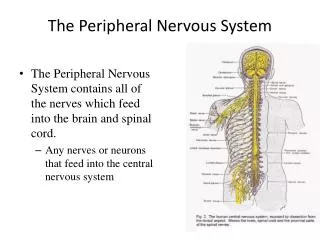
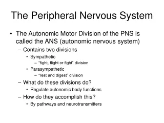
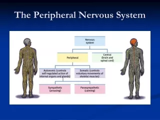
THE PERIPHERAL NERVOUS SYSTEM • PNS: includes all neural structures outside the brain and spinal cord, that is, the sensory receptors, peripheral nerves, and their associated ganglia, and efferent motor endings
SENSORY RECEPTORS • Sensory receptors are specialized to respond to changes in their environment called stimuli • Sensation: awareness of the stimulus (PNS) • Perception: interpretation of the meaning of the stimulus (CNS: brain)
SENSORY RECEPTORS • Sensory receptors may be classified 3 ways: • Receptors my be classified according to the activating stimulus • Receptors may be classified based on their location or the location of the activating stimulus • Receptors may be classified based on their overall structural complexity
SENSORY RECEPTORS • Classification based on Stimulus Type:overstimulation of anyone of the following receptors can result in pain: • Mechanoreceptors: generates nerve impulses when stimulated by forces (touch, pressure, vibration, stretch, itch) • Thermoreceptors: sensitive to temperature changes • Photoreceptors: respond to light energy (e.g. retina of the eye) • Chemoreceptors: respond to chemicals in solution (molecules smelled or tasted, or changes in blood chemistry) • Nociceptors: respond to potentially damaging stimuli that result in pain
SENSORY RECEPTORS • Classification based on Location: • Exteroceptors: • Sensitive to stimuli arising outside the body • Receptors near or at the body surface • Interoceptors: • Respond to stimuli arising from within the body • Internal viscera and blood vessels • Propriceptors: • Respond to internal stimuli from musculoskeletal organs • Skeletal muscle, joints, tendons, ligaments, connective tissue coverings of bone and muscle • Monitors degree of stretch of the organs
SENSORY RECEPTORS • Classification based on Complexity: • Simple: • Modified dendritic endings of sensory neurons • Found in the skin, mucous membranes, muscles, and connective tissues • Monitor most types of general sensory information • Complex: • Actually sense organs • Localized collection of cells working together to accomplish a specific receptive process (vision, hearing, smell, and taste)
SENSORY RECEPTORS • Anatomically: • Classified as free dendritic endings or encapsulated dendritic endings
SENSORY RECEPTORS • Free Dendritic Endings: • Naked • Present nearly everywhere in the body, but abundant in epithelia and connective tissue • Most unmyelinated • Small diameters • Free, or naked, nerve endings are present everywhere in the body and respond primarily to pain and temperature • Merkel discs: deep layers of skin epidermis • Function as light touch receptors • Root Hair Plexuses: light touch receptors that detect bending of hairs • Itch receptors: seemed to be stimulated by chemicals (histamine and bradykinin) present at inflamed sites
SENSORY RECEPTORS • Encapsulated Dendritic Endings: consist of one or more fiber terminals of sensory neurons enclosed in a connective tissue capsule • Virtually all are mechanoreceptors • Meissner’s corpuscles are receptors for discriminatory and light touch in hairless areas of the body • Tactile corpuscles • Skin • Krause’s end bulbs: variation of Meissner’s corpuscles • Abundant in mucous membranes • Mucocutaneous corpuscles • Pacinian, or lamellated, corpuscles, are stimulated when deep pressure is first applied • Deep in epidermis • Respond only when pressure is first applied • Ruffini’s corpuscles respond to deep and continuous pressure • Dermis • Muscle spindles detect when a muscle is being stretched and initiate a reflex that resists the stretch • Neuromuscular spindles • Golgi tendon organs are stimulated when the associated muscle stretches the tendon • Proprioceptors • Joint kinesthetic receptors monitor the stretch in the articular capsules of synovial joints
OVERVIEW: FROM SENSATION TO PERCEPTION • The somatosensory system:the part of the sensory system serving the body wall and limbs, involve the receptor level, the circuit level, and the perceptual level • Processing at the receptor level involves a stimulus that must excite a receptor in order for sensation to occur • Processing at the circuit level is involved with delivery of impulses to the appropriate region of the cerebral cortex for stimulus localization and perception • Processing at the perceptual level involves interpretation of sensory input in the cerebral cortex
NERVES AND ASSOCIATED GANGLIA • A nerve is a cordlike organ consisting of parallel bundles of peripheral axons (some myelinated and some not) enclosed by connective tissue wrappings: • Most mixed: both sensory and motor • Some are only sensory • Some are only motor • Ganglia are collections of neuron cell bodies associated with nerves in the PNS: • Ganglia associated with afferent nerve fibers contain cell bodies of sensory neurons • Ganglia associated with efferent nerve fibers mostly contain cell bodies of autonomic motor neurons
NERVE REGENERATION • Mature neurons do not divide • If damage to a neuron occurs to the axon and the cell body remains intact, cut or compressed axons can regenerate: • Post-trauma axon regrowth is never exactly the same as what existed before the injury • Much of the functional recovery after nerve injury involves retraining the nervous system to respond appropriately so that stimulus and response are coordinated • Unlike peripheral nerve fibers, most of those within the CNS never regenerate under normal circumstances (damage to the brain or spinal cord has been viewed as irreversible) • The key to CNS regeneration will most likely be found in the hippocampus, a brain region important in learning and memory because this area produces significant numbers of new neurons throughout life
NERVE REGENERATION • a.Nerve Regeneration: • Peripheral axon has been severed or crushed • Separated ends seal themselves off • Substances being transported along the axon begin to accumulate in the sealed ends
NERVE REGENERATION • b.Nerve Regeneration: • Within a few hours, the axon and its myelin sheath distal to the site of injury begin to disintegrate because they cannot receive nutrients (Wallerian degeneration) • Spreads distally from the injury site, completely fragmenting the axon • Macrophages phagocytize disintegrating myelin and axonal debris
NERVE REGENERATION • c.Nerve Regeneration: • Once the debris has been disposed of, surviving Schwann cells proliferate • Axonal growth takes place
NERVE REGENERATION • d.Nerve Regeneration: • The Schwann cells protect, support, and remyelinate the regenerating axons
NERVE REGENERATION • The greater the distance between the severed nerve endings, the less the chance of recovery because adjacent tissues block growth by protruding into the larger gaps • Neurosurgeons align cut nerve endings surgically to enhance the chance of successful regeneration (silicon tubes filled with biodegradable collagen) • The post-trauma axon regrowth is never exactly the same as what existed before the injury– pinpoint accuracy in nerve fibers (realignment) is impossible • Much of the functional recovery after nerve injury involves retraining the nervous system to respond appropriately so that stimulus and responses are coordinated
CRANIAL NERVES • Twelve pairs of cranial nerves are associated with the brain and pass through various foramina of the skull • The first two pairs attach to the forebrain; the rest originate from the brain stem • Other than the vagus nerves, which extend well into the abdomen, the cranial nerves serve only head and neck structures • Names reveal the most important structures they serve or their primary functions • Traditionally they are numbered using Roman Numerals
CRANIAL NERVES • Twelve pairs of cranial nerves are associated with the brain and pass through various foramina of the skull • The first two pairs attach to the forebrain; the rest originate from the brain stem • Other than the vagus nerves, which extend well into the abdomen, the cranial nerves serve only head and neck structures • Names reveal the most important structures they serve or their primary functions • Traditionally they are numbered using Roman numerals
CRANIAL NERVES • I: Olfactory nerves are responsible for smell • II: Optic nerves are responsible for vision • III: Oculomotor nerves play a role in eye movement • IV: Trochlear nerves play a role in eye movement • V: Trigeminal nerves are general sensory nerves of the face • VI: Abducens nerves play a role in eye movement • VII: Facial nerves function as the chief motor nerves of the face • VIII: Vestibulocochlearnerves are responsible for hearing and equilibrium • IX: Glossopharyngeal nerves innervate part of the tongue and pharynx • X: Vagus nerves innervate the heart, lungs, and the abdominal organs • XI: Accessory nerves move structures associated with the head and neck • XII: Hypoglossal nerves are mixed nerves that arise from the medulla and serve the tongue
CRANIAL NERVES • I: Olfactory nerves are responsible for smell • II: Optic nerves are responsible for vision • III: Oculomotor nerves play a role in eye movement • IV: Trochlear nerves play a role in eye movement • V: Trigeminal nerves are general sensory nerves of the face • VI: Abducens nerves play a role in eye movement • VII: Facial nerves function as the chief motor nerves of the face • VIII: Vestibulocochlear nerves are responsible for hearing and equilibrium • IX: Glossopharyngeal nerves innervate part of the tongue and pharynx • X: Vagus nerves innervate the heart, lungs, and the abdominal organs • XI: Accessory nerves move structures associated with the head and neck • XII: Hypoglossal nerves are mixed nerves that arise from the medulla and serve the tongue
CRANIAL NERVES • On occasion, our trusty truck acts funny– very good vehicle anyhow
On occasion, our trusty truck acts funny– very good vehicle anyhow I: Olfactory nerves are responsible for smell II: Optic nerves are responsible for vision III: Oculomotor nerves play a role in eye movement IV: Trochlear nerves play a role in eye movement V: Trigeminal nerves are general sensory nerves of the face VI: Abducens nerves play a role in eye movement VII: Facial nerves function as the chief motor nerves of the face VIII: Vestibulocochlear nerves are responsible for hearing and equilibrium IX: Glossopharyngeal nerves innervate part of the tongue and pharynx X: Vagus nerves innervate the heart, lungs, and the abdominal organs XI: Accessory nerves move structures associated with the head and neck XII: Hypoglossal nerves are mixed nerves that arise from the medulla and serve the tongue CRANIAL NERVES
CRANIAL NERVES • I II III IV V VI VII • Some say marry money, but my brother • VIII IX X XI XII • says (its) bad business (to) marry money
CRANIAL NERVES • I II III IV V VI VII • Some say marry money, but my brother • VIII IX X XI XII • says (its) bad business (to) marry money
SPINAL NERVES • Thirty-one pairs of mixed spinal nerves arise from the spinal cord and serve the entire body except the head and some areas of the neck • All are mixed nerves • Named according to their point of issue from the spinal cord • 8 pairs of cervical spinal nerves (C1 – C8) • 12 pairs of thoracic nerves (T1 – T12) • 5 pairs of lumbar nerves (L1 – L5) • 5 pairs of sacral nerves (S1 – S5) • 1 pair of tiny coccygeal nerves ( C0) • Notice that there are 8 pairs of cervical nerves but only 7 cervical vertebrae • First 7 pairs of cervical nerves exit the vertebral canal superior to the vertebrae for which they are named (C8 emerges inferiorto the 7th cervical vertebra– between C7 and T1) • All the remaining spinal nerves leave the vertebral column inferior to the same-numbered vertebra
SPINAL NERVES • Innervation of Specific Body Regions: • Each spinal nerve connects to the spinal cord by a dorsal root and a ventral root • Rami lie distal to and are lateral branches of the spinal nerves that carry both motor and sensory fibers • The back is innervated by the dorsal rami with each rami innervating the muscle in line with the point of origin from the spindle column • The cervical plexus is formed by the ventral rami of the first four cervical nerves • The brachial plexus is situated partly in the neck and partly in the axilla and gives rise to virtually all the nerves that innervate the upper limb • Only in the thorax are the ventral rami arranged in a simple segmental pattern corresponding to that of the dorsi rami • The area of skin innervated by the cutaneous branches of a single nerve is called a dermatone • Hinton’s law states that any nerve serving a muscle that produces movement at a joint also innervates the joint and the skin over the joint
SPINAL NERVES • Innervation of Specific Body Regions: • Each spinal nerve connects to the spinal cord by a dorsal root and a ventral root • Rami lie distal to and are lateral branches of the spinal nerves that carry both motor and sensory fibers
SPINAL NERVES • The cervical plexus is formed by the ventral rami of the first four cervical nerves • The brachial plexus is situated partly in the neck and partly in the axilla and gives rise to virtually all the nerves that innervate the upper limb • Only in the thorax are the ventral rami arranged in a simple segmental pattern corresponding to that of the dorsi rami • The sacral and lumbar plexuses overlap and because many fibers of the lumber plexus contribute to the sacral plexus via the lumbosacral trunk, the two plexuses are often referred to as the lumbosacral plexus • The area of skin innervated by the cutaneous branches of a single nerve is called a dermatone • Hinton’s law states that any nerve serving a muscle that produces movement at a joint also innervates the joint and the skin over the joint
FORMATION OF SPINAL NERVES • Formation of one pair of spinal nerves by the union of the ventral and dorsal roots of the spinal cord
FORMATION OF SPINAL NERVES • Spinal nerve is very short • Roots lie medial to and form the spinal nerves • Each root is strictly sensory or motor in function • Ramus(branch of a nerve, artery, vein, or bone) lie distal to and are lateral branches of the spinal nerves • Like spinal nerves, carry both sensory and motor fibers
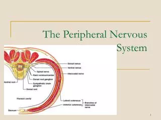
RAMI DISTRIBUTION OF THE SPINAL NERVE • Cross-sectional view of the left side of the body at the level of the thorax, showing the distribution of the dorsal and ventral rami of the spinal nerve • Notice the rami communicantes branches of the spinal nerve • Spinal nerve is very short • Emerging from its foramen it divides into the dorsal, ventral, and meningeal branches (reenters the vertebral canal to innervate the meninges and blood vessels within)
PLEXUS • A network of converging and diverging nerve fibers, blood vessels, or lymphatics
CERVICAL PLEXUS • Single most important nerve from this plexus is the phrenic nerve • Input from C3 and C4 • Supplies both motor and sensory fibers to the diaphragm which is a major muscle in the breathing • Irritation of this nerve causes spasms resulting in hiccups • If severed, the diaphragm is paralyzed and respiration arrest occurs • Kept alive by mechanical respirators
BRACHIAL PLEXUS • Gives rise to virtually all the nerves that innervate the upper limb • Rami (roots) gives rise to trunks then divisions then cords (Really Tired Drink Coffee) • Cords are namedfor their relationship to the axillary artery (lateral / posterior / medial) • The three cords gives rise to the main nerves of the upper limb: • Axillary (deltoid/teres major/skin/shoulder joint) • Musculocutaneous (biceps brachii/brachialis/skin) • Median (skin/most flexors/lateral palm) • Ulnar (medial hand/intrinsic hand/finger and wrist flexion) • Radial: largest branch (extensors) • Nerve rest against the medial epicondyle of the humerus (funny bone) • Striking results in tingling of the little finger • Compression and ischemia (decrease blood supply) can result in temporary paralysis