THE PERIPHERAL NERVOUS SYSTEM AND REFLEX ACTIVITY
470 likes | 2.39k Views
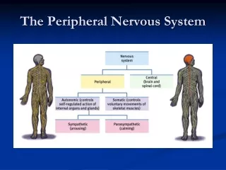
THE PERIPHERAL NERVOUS SYSTEM AND REFLEX ACTIVITY. PNS in the Structural Organization of the Nervous system. SENSORY RECEPTORS AND SENSATION. SENSORY RECEPTORS. Sensory receptors are specialized to respond to changes in their environment called stimuli

THE PERIPHERAL NERVOUS SYSTEM AND REFLEX ACTIVITY
E N D
Presentation Transcript
SENSORY RECEPTORS • Sensory receptors are specialized to respond to changes in their environment called stimuli • Receptors my be classified according to the activating stimulus • Receptors may be classified based on their location or the location of the activating stimulus • Receptors may be classified based on their overall structural complexity
SENSORY RECEPTORS • Free, or naked, nerve endings are present everywhere in the body and respond primarily to pain and temperature • Encapsulated Dendritic Endings • Meissner’s corpuscles are receptors for discriminatory and light touch in hairless areas of the body • Pacinian, or lamellated, corpuscles, are stimulated when deep pressure is first applied • Ruffini’s corpuscles respond to deep and continuous pressure • Muscle spindles detect when a muscle is being stretched and initiate a reflex that resists the stretch • Golgi tendon organs are stimulated when the associated muscle stretches the tendon • Joint kinesthetic receptors monitor the stretch in the articular capsules of synovial joints
OVERVIEW: FROM SENSATION TO PERCEPTION • The somatosensory system, the part of the sensory system serving the body wall and limbs, involve the receptor level, the circuit level, and the perceptual level • Processing at the receptor level involves a stimulus that must excite a receptor in order for sensation to occur • Processing at the circuit level is involved with delivery of impulses to the appropriate region of the cerebral cortex for stimulus localization and perception • Processing at the parceptual level involves interpretation of sensory input in the cerebral cortex
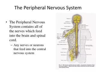
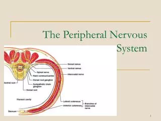
NERVES AND ASSOCIATED GANGLIA • A nerve is a cordless organ consisting of parallel bundles of peripheral axons enclosed by connective tissue wrappings • Ganglia are collections of neuron cell bodies associated with nerves in the PNS • If damage to a neuron occurs to the axon and the cell body remains intact, cut or compressed axons can regenerate
CRANIAL NERVES • Olfactory nerves are responsible for smell • Optic nerves are responsible for vision • Oculomotor nerves play a role in eye movement • Trochlear nerves play a role in eye movement • Trigeminal nerves are general sensory nerves of the face • Abducens nerves play a role in eye movement • Facial nerves function as the chief motor nerves of the face • Vestibulocochlear nerves are responsible for hearing and equilibrium • Glossopharyngeal nerves innervate part of the tongue and pharynx • Vagus nerves innervate the heart, lungs, and the abdominal organs • Accessory nerves move structures associated with the head and neck • Hypoglossal nerves are mixed nerves that arise from the medulla and serve the tongue
SPINAL NERVES • Thirty-one pairs of mixed spinal nerves arise from the spinal cord and serve the entire body except the head and neck • Innervation of Specific Body Regions • Each spinal nerve connects to the spinal cord by a dorsal root and a ventral root • Rami lie distal to and are lateral branches of the spinal nerves that carry both motor and sensory fibers • The back is innervated by the dorsal rami with each rami innervating the muscle in line with the point of origin from the spindle column • Only in the thorax are the ventral rami arranged in a simple segmental pattern corresponding to that of the dorsi rami • The cervical plexus is formed by the ventral rami of the first four cervical nerves • The brachial plexus is situated partly in the neck and partly in the axilla and gives rise to virtually all the nerves that innervate the upper limb • The sacral and lumbar plexuses overlap and because many fibers of the lumber plexus contribute to the sacral plexus via the lumbosacral trunk, the two plexuses are often referred to as the lumbosacral plexus • The area of skin innervated by the cutaneous branches of a single nerve is called a dermatone • Hinton’s law states that any nerve serving a muscle that produces movement at a joint also innervates the joint and the skin over the joint
PERIPHERAL MOTOR ENDINGS • Peripheral motor endings are the PNS element that activates effectors by releasing neurotransmitters • The terminals of the somatic motor fibers that innervate voluntary muscles form elaborate neuromuscular junctions with their effector cells and they release the neurotransmitter acetylcholine • The junctions between autonomic motor endings and the visceral effectors involve varicosities and release either acetylcholine or epinephrine as their neurotransmitter
LEVELS OF MOTOR CONTROL • The segmental level is the lowest level on the motor control hierarchy and consists of the spinal cord circuits • The projection level has direct control of the spinal cord • The precommand level is made up of the cerebellum and the basal nuclei and is the highest level of the motor system hierarchy
THE REFLEX ARC • Reflexes are unlearned, rapid, predictable motor responses to a stimulus, and occur over highly specific neural pathways called reflex arc
SPINAL REFLEXES • Spinal reflexes are somatic reflexes mediated by the spinal cord • In the stretch reflex the muscle spindle is stretched and excited by either an external stretch or an internal stretch • The Golgi tendon reflex produces muscle relaxation and lengthening in response to contraction • The flexor, or withdrawal, reflex is initiated by a painful stimulus and causes automatic withdrawal of the threatened body part from the stimulus • They crossed extensor reflex is a complex spinal reflex consisting of an ipsilateral withdrawal reflex and a contralateral extensor reflex • Superficial reflexes are elicited by gentle cutaneous stimulation
DEVELOPMENTAL ASPECTS OF THE PERIPHERAL NERVOUS SYTEM • The Spinal Nerves branch from the developing spinal cord and adjacent neural crest and exit between the forming vertebrate • Each nerve becomes associated with the adjacent muscle mass • Cranial Nerves innervate muscles of the head in a similar way • Sensory Receptors atrophy to some degree with age, and there is a decrease in muscle tone in the face and neck, reflexes occur a bit more slowly